Showing posts with label lupus rashes. Show all posts

Wednesday, January 29, 2014

Urticarial Vasculitis-It's a lupus thing!


It's a little difficult to see in the pic-but I tried to capture not the rash or hives on top of the skin but the bumps underneath the skin.  My blood vessels were so inflammed I could follow them up and down my arms-and feel the muscles and nerves of my ulnar and carpal neuropathy being pulled.

The actual term is urticarial vasculitis.  It's a hypersensitivity vascular reaction to the UV or the sun-of which many people with lupus have sensitivities with.  When mine gets bad-it causes this reaction.  I call it a mini-flare-it usually takes about a week to come all the way down, and yes, it ITCHES and yes, it's painful.  While I usually have it on my arms, I have had it on my chest and face as well.  Here's my chest, only slightly reactive, but still very hot, swollen and itchy:  Image
The discolorations you see on my chest are from previous urticaria rashes, lupus rashes, discoid rashes, lesions and pemphigoid rashes.  Here's some info on urticarial vasculitis from medscape:
Urticarial vasculitis is an eruption of erythematous wheals that clinically resemble urticaria but histologically show changes of leukocytoclastic vasculitis.[1, 2] Urticarial vasculitis may be divided into normocomplementemic and hypocomplementemic variants. Both subsets can be associated with systemic symptoms (eg, angioedema, arthralgias, abdominal or chest pain, fever, pulmonary disease, renal disease, episcleritis, uveitis, and Raynaud phenomenon).

The hypocomplementemic form more often is associated with systemic symptoms and has been linked to connective-tissue disease (ie, systemic lupus erythematosus [SLE]).

Patients with urticarial vasculitis present with an urticarial eruption, often accompanied by a painful or burning sensation. Lesions are generalized wheals or erythematous plaques, occasionally with central clearing, lasting for more than 24 hours in a fixed location (in contrast to urticaria, which resolves in minutes to hours or migrates continually). Petechiae may be noted within the lesions, and they may resolve with ecchymoses or postinflammatory hyperpigmentation. Patients may have photosensitivity, lymphadenopathy, arthralgia, angioedema (40%), fever, abdominal pain, dyspnea, and pleural and pericardial effusions.[4] Most cases of urticarial vasculitis are idiopathic.
The primary causes of urticarial vasculitis are as follows:
  • Drug induced, such as ACE inhibitors, penicillin, sulfonamides, fluoxetine, cimetidine, diltiazem, thiazides, potassium iodide, non-steroid inflammatory drugs, and glatiramer acetate.[9]
  • Rheumatic disease, such as SLE and Sjögren syndrome: Urticarial vasculitis has also been reported with immunoglobulin A and immunoglobulin M monoclonal gammopathies, mixed cryoglobulins, and hematologic and solid malignancies.[10]
Urticarial vasculitis is divided into hypocomplementemic and normocomplementemic categories, as follows[12] :
  • Hypocomplementemia often is associated with a systemic condition, such as SLE (in which >50% of patients have hypocomplementemia).[3] In addition, as many as 71% of patients with hypocomplementemic urticarial vasculitis have a positive antinuclear antibody titer but do not fulfill the American Rheumatism Association criteria for SLE.[5] Some authors have suggested evaluation of hypocomplementemic urticarial vasculitis for immunoglobulin G antibodies to C1q. Individuals with these antibodies have a higher incidence of angioedema, ocular inflammation, glomerulonephritis, and obstructive pulmonary disease.
  • Normocomplementemic vasculitis can be associated with connective-tissue disease but at a much lower rate.

Sunday, May 26, 2013

Learning to move on from where I am and not from where I was!

I couldn’t pick up where I left off before my last bout with the sun and sun caused illness.
Slowly moving in and out of social networks, commenting, reading emails, making and keeping doctor appointments, getting out and all in all joining in my own life as a contributor and not a spectator takes all my energy and thought processes.  Catching up is too hard so I’m learning to just move on, click the like button on fb on new posts and put recent sicknesses that stopped me in their tracks behind me.  This will be my only mention of it and I’m keeping it light.

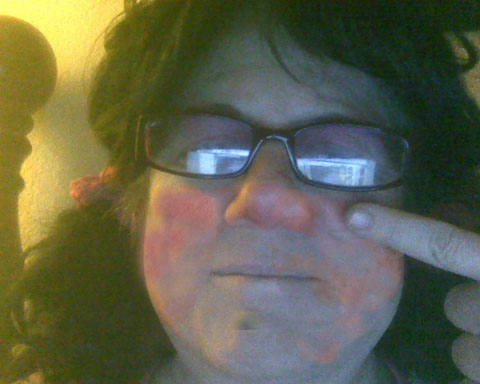
I had what they called a hypersensitive vasculitis facial attack from the sun (UV).  I wasn’t just down for the count, I was blasted into an oblivion of pain like even I haven’t experienced in quite a long time. My face and arm itched from the inside and burned as if my blood was boiling from the inside.  After the hives disappeared and the bleeding stopped my face on and under my lupus malar rash became HOT and SWOLLEN.  I looked like someone punched me.  My nerves and every layer of skin were on fire.  I went 3 days until it dissipated enough with steroids that I could talk without searing pain.  A tonage of anti-inflammatories and 60mg of the dreaded prednisone later the swelling went down and the burning under my skin subsided.  I NEVER EVER AGAIN want to go through this.  I’m guessing the appearance of the newly summerized hot Arizona sun and UV in the danger levels precipitated the attack.  Right now I think I’d like to just stay inside the rest of my life.  But I won’t.  Who would?
juliesunmay2013
Here’s a little info on hypersensitive vasculitis from medscape.com:

Hypersensitivity vasculitis, which is usually represented histopathologically as leukocytoclastic vasculitis, is a term commonly used to denote a small vessel vasculitis. Many possible causes or associations exist for hypersensitivity vasculitis, but a cause or an associated disorder is not found in as many as 50% of patients.
Hypersensitivity vasculitis (a form of small vessel vasculitis) may manifest clinically as cutaneous disease only or it may manifest as skin disease with involvement of other organs. The internal organs most commonly affected in hypersensitivity vasculitis are the joints, gastrointestinal tract, and the kidneys. The prognosis for hypersensitivity vasculitis is good when no internal involvement is clinically present. Hypersensitivity vasculitis may be acute and self-limited, recurrent or chronic.

Patients with hypersensitivity vasculitis of their skin may report itching, a burning sensation, or pain, or they may have asymptomatic lesions. Vasculitis of the skin may occur in the absence of any detectable systemic disease. Vasculitis may occur in conjunction with collagen-vascular disorders, paraproteinemia, ingestants (drugs or foods), infections, or malignancy (rare).

I’m prone to severe sun reactions, pemphigoid rashes, porphyric reactions, discoid rashes, hives, necrotizing lesions, livedo reticularis, small spot vasculitis, reynauds, but this is something different.  This is cellular pain (like cellulitis) that effects you down to the core.  It’s horrific.  I assure you this autoimmune reaction is not a sunburn.  I wish it was.  Boy do I.

My body is recovering.  My mind is fine.  I’m not picking up where I left off anymore.  I’m moving on.  Life is too short and too precious.  HUGS!

Tuesday, April 23, 2013

Is it Shingles or one of my Cutaneous Porphyria rashes?


It's ITCHY AS HECK!
It's HOT HOT HOT and inflammed.
It "pocked up" in a day
It followed a nerve path
It happened after a cold sore attack
Was in the sun & have LUPUS & Cutaneous Porphyria

What do YOU think?
I need to plan a viral test WHEN I get this rash.  Did I mention it was ITCHY?

To Explain the photosensitive process in the skin, I have systemic lupus and with that circulating immune complexes in my blood that do the actual damage to tissue and skin.  Here is an explanation of that hypersensitve process:  Not all my rashes are necrotizing, but many are.
from http://en.wikipedia.org/wiki/Type_III_hypersensitivity
Type III hypersensitivity occurs when antigen-antibody complexes that are not adequately cleared by innate immune cells accumulate, giving rise to an inflammatory response and attraction of leukocytes.

Presentation

Type III hypersensitivity occurs when there is little antigen and an excess of antibody, leading to small immune complexes being formed that do not fix complement and are not cleared from the circulation. It is characterized by solvent antigens that are not bound to cell surfaces (which is the case in type II hypersensitivity). When these antigens bind antibodies, immune complexes of different sizes form.[1] Large complexes can be cleared by macrophages but macrophages have difficulty in the disposal of small immune complexes. These immune complexes insert themselves into small blood vessels, joints, and glomeruli, causing symptoms. Unlike the free variant, a small immune complex bound to sites of deposition (like blood vessel walls) are far more capable of interacting with complement; these medium-sized complexes, formed in the slight excess of antigen, are viewed as being highly pathogenic.[2]
Such depositions in tissues often induce an inflammatory response,[3] and can cause damage wherever they precipitate. The cause of damage is as a result of the action of cleaved complement anaphylotoxins C3a and C5a, which, respectively, mediate the induction of granule release from mast cells (from which histamine can cause urticaria), and recruitment of inflammatory cells into the tissue (mainly those with lysosomal action, leading to tissue damage through frustrated phagocytosis by PMNs and macrophages).[4]
The reaction can take hours, days, or even weeks to develop, depending on whether or not there is immunlogic memory of the precipitating antigen. Typically, clinical features emerge a week following initial antigen challenge, when the deposited immune complexes can precipitate an inflammatory response. Because of the nature of the antibody aggregation, tissues that are associated with blood filtration at considerable osmotic and hydrostatic gradient (e.g. sites of urinary and synovial fluid formation, kidney glomeruli and joint tissues respectively) bear the brunt of the damage. Hence, vasculitis, glomerulonephritis and arthritis are commonly-associated conditions as a result of type III hypersensitivity responses[5]).
As observed under methods of histopathology, acute necrotizing vasculitis within the affected tissues is observed concomitant to neutrophilic infiltration, along with notable eosinophilic deposition (fibrinoid necrosis). Often, immunofluorescence microscopy can be used to visualize the immune complexes [5]). Skin response to a hypersensitivity of this type is referred to as an Arthus reaction, and is characterized by local erythema and some induration. Platelet aggregation, especially in microvasculature, can cause localized clot formation, leading to blotchy hemorrhages. This typifies the response to injection of foreign antigen sufficient to lead to the condition of serum sickness.[4]

Tuesday, February 5, 2013

Pemphigus Erythematosus or AKA lupus discoid rash with weeping

First my rash.  I saw my rheumie and he said something about it being pemphigus.  I thought it was just subacute cutaneous lupus.  (in addition to my SLE)  Btw I am very confused about lupus rashes, lol. 

Here is a pic of the rash and then another pic of it healing five days later.  It itched, it seeped it was in a circle with dark edges.  It hurt and pulled the little hairs on my neck.  I"d wake up with my long hair stuck to my neck.  Really gross stuff.  I had one like this on my chest (left me discolored but not scarred) and one on my face and one on my head.  (see my rashes page).


The various types of pemphigus include pemphigus erythematosus, pemphigoid, pemphigus vegetans, pemphigus vulgaris, and pemphigus foliaceus.

Pemphigus erythematosus, also known as Senear-Usher syndrome, is an overlap syndrome with features of lupus erythematosus (LE) and pemphigus foliaceus. Pemphigus is demonstrated by acantholysis and immunoglobulin deposits in the interkeratinocyte substance (see the image below).
Direct immunofluorescence microscopy performed on Direct immunofluorescence microscopy performed on epithelial biopsy specimen obtained from a patient with pemphigus vulgaris detects immunoglobulin G deposits at the epithelial cell surfaces. 
 The lupus component of pemphigus erythematosus is demonstrated by circulating antinuclear antibodies (ANA) and sometimes by immunoglobulin and complement deposits at the dermoepidermal junction
For a thorough description and introduction to the possible causes of pemphigus, see the article "Pemphigus: An Acronym for a Disease with Multiple Causes ", published by the International Pemphigus Society.[1]