Thursday, April 3, 2014

Get the SKIN-EEEE on Scleroderma!

Image
Scleroderma=Sclera=hardening
There are two types, diffuse or skin.  My scleroderma is limited to my skin.  I’m VERY lucky! (even if I don’t look it in the middle pic, lol)  You may have to click on the pic to get a close up of it so you can really tell how thick and “pulled” the skin is.  Yes, it was painful.  It took about two weeks to go “down” and get back to normal.  I used steroid cream to heal and prednisone.  It was the worse rash of it’s kind on my face I ever had.  The pic on the right shows the hardening and peeling fingertips common in Scleroderma-called “scleroderma fingers”.

Here’s some more info on Scleroderma from Scleroderma.org:
Scleroderma, or systemic sclerosis, is a chronic connective tissue disease generally classified as one of the autoimmune rheumatic diseases.

The word “scleroderma” comes from two Greek words: “sclero” meaning hard, and “derma” meaning skin. Hardening of the skin is one of the most visible manifestations of the disease. The disease has been called “progressive systemic sclerosis,” but the use of that term has been discouraged since it has been found that scleroderma is not necessarily progressive. The disease varies from patient-to-patient.

What scleroderma is not
Scleroderma is not contagious, infectious, cancerous or malignant.
How serious is scleroderma?
Any chronic disease can be serious. The symptoms of scleroderma vary greatly for each person, and the effects of scleroderma can range from very mild to life threatening. The seriousness will depend on the parts of the body, which are affected, and the extent to which they are affected. A mild case can become more serious if not properly treated. Prompt and proper diagnosis and treatment by qualified physicians may minimize the symptoms of scleroderma and lessen the chance for irreversible damage.

How is scleroderma diagnosed?
The diagnostic process may require consultation with rheumatologists (arthritis specialists), and/or dermatologists (skin specialists) and require blood studies and numerous other specialized tests depending upon which organs are affected.

Who develops scleroderma, and when?
It’s estimated that about 300,000 Americans have scleroderma. About one third of those people have the systemic form of scleroderma. Since scleroderma presents with symptoms similar to other autoimmune diseases, diagnosis is difficult. There may be many misdiagnosed or undiagnosed cases.
Localized scleroderma is more common in children, whereas systemic scleroderma is more common in adults. Overall, female patients outnumber male patients about 4-to-1. Factors other than a person’s gender, such as race and ethnic background, may influence the risk of getting scleroderma, the age of onset, and the pattern or severity of internal organ involvement. The reasons for this are not clear. Although scleroderma is not directly inherited, some scientists feel there is a slight predisposition to it in families with a history of rheumatic diseases.

However, scleroderma can develop in every age group from infants to the elderly, but its onset is most frequent between the ages of 25 to 55. When doctors say “usually” or “for the most part,” the reader should understand that variations frequently occur. Many patients get alarmed when they read medical information that seems to contradict their own experiences, and conclude that what has happened to them is not supposed to happen. There are many exceptions to the rules in scleroderma, perhaps more so than in other diseases. Each case is different, and information should be discussed with your own doctor.

What causes scleroderma?
The exact cause or causes of scleroderma are still unknown, but scientists and medical researchers are working hard to make those determinations. It is known that scleroderma involves an overproduction of collagen.

Is scleroderma genetic?
Most patients do not have any relatives with scleroderma and their children do not get scleroderma. Research indicates that there is a susceptibility gene, which raises the likelihood of getting scleroderma, but by itself does not cause the disease.

What is the treatment for scleroderma?
Currently, there is no cure for scleroderma, but there are many treatments available to help particular symptoms. For instance, heartburn can be controlled by medications called proton pump inhibitors PPIs) or medicine to improve the motion of the bowel. Some treatments are directed at decreasing the activity of the immune system. Some people with mild disease may not need medication at all and occasionally people can go off treatment when their scleroderma is no longer active. Because there is so much variation from one person to another, there is great variation in the treatments prescribed.

Types of Scleroderma

There are two major classifications of scleroderma: localized scleroderma and systemic sclerosis (SSc). Other forms or subclassifications, each with its own characteristics and prognosis, may be identified through future research.
types of scleroderma chart
Localized Scleroderma The changes, which occur in localized scleroderma, are usually found in only a few places on the skin or muscles, and rarely spread elsewhere. Generally, localized scleroderma is relatively mild. The internal organs are usually not affected, and persons with localized scleroderma rarely develop systemic scleroderma. Some laboratory abnormalities commonly seen in systemic scleroderma are frequently absent in the localized form.
Morphea is a form of localized scleroderma characterized by waxy patches on the skin of varying sizes, shapes and color. The skin under the patches may thicken. The patches may enlarge or shrink, and often may disappear spontaneously. Morphea usually appears between the ages of 20 and 50, but is often seen in young children.
Linear scleroderma is a form of localized scleroderma which frequently starts as a streak or line of hardened, waxy skin on an arm or leg or on the forehead. Sometimes it forms a long crease on the head or neck, referred to as en coup de sabre because it resembles a saber or sword wound. Linear scleroderma tends to involve deeper layers of the skin as well as the surface layers, and sometimes affects the motion of the joints, which lie underneath. Linear scleroderma usually develops in childhood. In children, the growth of involved limbs may be affected.
Systemic scleroderma (systemic sclerosis) The changes occurring in systemic scleroderma may affect the connective tissue in many parts of the body. Systemic scleroderma can involve the skin, esophagus, gastrointestinal tract (stomach and bowels), lungs, kidneys, heart and other internal organs. It can also affect blood vessels, muscles and joints. The tissues of involved organs become hard and fibrous, causing them to function less efficiently. The term systemic sclerosis indicates that “sclerosis” (hardening) may occur in the internal systems of the body. There are two major recognized patterns that the illness can take – diffuse or limited disease. In diffuse scleroderma, skin thickening occurs more rapidly and involves more skin areas than in limited disease. In addition, people with diffuse scleroderma have a higher risk of developing “sclerosis” or fibrous hardening of the internal organs.
About 50 percent of patients have a slower and more benign illness called limited scleroderma. In limited scleroderma, skin thickening is less widespread, typically confined to the fingers, hands and face, and develops slowly over years. Although internal problems occur, they are less frequent and tend to be less severe than in diffuse scleroderma, and are usually delayed in onset for several years. However, persons with limited scleroderma, and occasionally those with diffuse scleroderma,
can develop pulmonary hypertension, a condition in which the lung’s blood vessels become narrow, leading to impaired blood flow through the lungs resulting in shortness of breath.
Limited scleroderma is sometimes called CREST syndrome. CREST stands for the initial letters of five common features:
  • Calcinosis
  • Raynaud Phenomenon
  • Esophageal dysfunction
  • Sclerodactyly
  • Telangiectasia
To further complicate the terminology, some people with diffuse disease will go on to develop calcinosis and telangiectasias so that they also have the features of CREST.
Although most patients can be classified as having diffuse or limited disease, different people may have different symptoms and different combination of symptoms of the illness.

Wednesday, January 29, 2014

Urticarial Vasculitis-It's a lupus thing!


It's a little difficult to see in the pic-but I tried to capture not the rash or hives on top of the skin but the bumps underneath the skin.  My blood vessels were so inflammed I could follow them up and down my arms-and feel the muscles and nerves of my ulnar and carpal neuropathy being pulled.

The actual term is urticarial vasculitis.  It's a hypersensitivity vascular reaction to the UV or the sun-of which many people with lupus have sensitivities with.  When mine gets bad-it causes this reaction.  I call it a mini-flare-it usually takes about a week to come all the way down, and yes, it ITCHES and yes, it's painful.  While I usually have it on my arms, I have had it on my chest and face as well.  Here's my chest, only slightly reactive, but still very hot, swollen and itchy:  Image
The discolorations you see on my chest are from previous urticaria rashes, lupus rashes, discoid rashes, lesions and pemphigoid rashes.  Here's some info on urticarial vasculitis from medscape:
Urticarial vasculitis is an eruption of erythematous wheals that clinically resemble urticaria but histologically show changes of leukocytoclastic vasculitis.[1, 2] Urticarial vasculitis may be divided into normocomplementemic and hypocomplementemic variants. Both subsets can be associated with systemic symptoms (eg, angioedema, arthralgias, abdominal or chest pain, fever, pulmonary disease, renal disease, episcleritis, uveitis, and Raynaud phenomenon).

The hypocomplementemic form more often is associated with systemic symptoms and has been linked to connective-tissue disease (ie, systemic lupus erythematosus [SLE]).

Patients with urticarial vasculitis present with an urticarial eruption, often accompanied by a painful or burning sensation. Lesions are generalized wheals or erythematous plaques, occasionally with central clearing, lasting for more than 24 hours in a fixed location (in contrast to urticaria, which resolves in minutes to hours or migrates continually). Petechiae may be noted within the lesions, and they may resolve with ecchymoses or postinflammatory hyperpigmentation. Patients may have photosensitivity, lymphadenopathy, arthralgia, angioedema (40%), fever, abdominal pain, dyspnea, and pleural and pericardial effusions.[4] Most cases of urticarial vasculitis are idiopathic.
The primary causes of urticarial vasculitis are as follows:
  • Drug induced, such as ACE inhibitors, penicillin, sulfonamides, fluoxetine, cimetidine, diltiazem, thiazides, potassium iodide, non-steroid inflammatory drugs, and glatiramer acetate.[9]
  • Rheumatic disease, such as SLE and Sjögren syndrome: Urticarial vasculitis has also been reported with immunoglobulin A and immunoglobulin M monoclonal gammopathies, mixed cryoglobulins, and hematologic and solid malignancies.[10]
Urticarial vasculitis is divided into hypocomplementemic and normocomplementemic categories, as follows[12] :
  • Hypocomplementemia often is associated with a systemic condition, such as SLE (in which >50% of patients have hypocomplementemia).[3] In addition, as many as 71% of patients with hypocomplementemic urticarial vasculitis have a positive antinuclear antibody titer but do not fulfill the American Rheumatism Association criteria for SLE.[5] Some authors have suggested evaluation of hypocomplementemic urticarial vasculitis for immunoglobulin G antibodies to C1q. Individuals with these antibodies have a higher incidence of angioedema, ocular inflammation, glomerulonephritis, and obstructive pulmonary disease.
  • Normocomplementemic vasculitis can be associated with connective-tissue disease but at a much lower rate.

Saturday, January 18, 2014

What's Phenomenal about Raynaud's Phenomenon?

juliehandsnewIt's winter, it's cold and that means people with autoimmune conditions can have an increase in symptoms, like joint pain, arthritis inflammation and raynaud's syndrome to name a few.
Raynauds is when small arteries that provide blood to your skin, narrow. This can cause trouble in circulation. In some people, it can turn your fingertips blue, purple or even white. Raynaud's can be minor-like in mine (pic) or so complicated it can cause amputations to be needed.
Raynaud's is more than cold hands-it can be very difficult to get your hands or feet to warm up after a flare. Preventative care is to layer up in clothes and wear nice warm socks and gloves.
Treatment according to Mayo.com:
Medications
Depending on the cause of your symptoms, medications may help treat Raynaud's. To widen (dilate) blood vessels and promote circulation, your doctor may prescribe:
Calcium channel blockers. These drugs relax and open up small blood vessels in your hands and feet. They decrease the frequency and severity of attacks in most people with Raynaud's. These drugs can also help heal skin ulcers on your fingers or toes. Examples include nifedipine (Adalat CC, Afeditab CR, Procardia), amlodipine (Norvasc) and felodipine (Plendil).
Alpha blockers. Some people find relief with drugs called alpha blockers, which counteract the actions of norepinephrine, a hormone that constricts blood vessels. Examples include prazosin (Minipress) and doxazosin (Cardura).
Vasodilators. Some doctors prescribe a vasodilator — a drug that relaxes blood vessels — such as nitroglycerin cream to your fingers to help heal skin ulcers. Your doctor may also prescribe vasodilator drugs that are commonly used to treat other conditions, but may effectively relieve the symptoms of Raynaud's. These drugs include the high blood pressure drug losartan (Cozaar), the erectile dysfunction medication sildenafil (Viagra, Revatio), the antidepressant medication fluoxetine (Prozac, Sarafem), and a class of medication called prostaglandins.

Friday, October 25, 2013

The Nerve of THIS NERVE-Failed Amputation-Going for Round 3!

The #NERVE of THIS NERVE; #Amputation Failed! Not giving up!
Yesterday I saw my surgeon-the amputation was a fail. The darn median nerve continues to curse my days and sleep. It went five days nonstop. Doc gave me no less than EIGHT lidocaine shots yesterday to shut it down. By the time he was done I couldn't feel any part of my right foot EXCEPT THE NERVE PAIN. I mean, it practically defies nature. The other possibility according to the million dollar nerve conduction equipment is that there are severely damaged small to medium fiber peripheral nerves in the skin that was used to close up the amputation.

Anyway, THIS IS WAR (as my BIL used to say)

End of story is I'm going in for another surgery. I'll let you know when it is. He's cutting the median nerve in two new places and ripping out the skin used to cover up where the toe is amputated and stretching what's left to use. He may have to graft later if it doesn't take and I get a wound problem. Ah, another risk I'm willing to take-wound problems. Like I said, "THIS IS WAR" and I'm not backin down until this nerve is gone for good.

I know what your thinking but I'm actually very lucky to have a surgeon willing to go thrice trying to help me. This is his last suggestion, after that-he is outta ideas. You've got to remember I am a complicated case with circulating immune complexes causing inflammation in my blood vessels that press on nerves and damage them. This is my particular present from lupus.
So.......that's whats happening. :)

Tuesday, September 17, 2013

Toe Amputated-Goodbye Neuroma! Hello Successful SSDI Hearing! Goodbye Lidocaine Overdose!


So much has happened! I’ll break it up in Parts.  Part 1-toe amputation.  Part 2- I WON at my disability appeal hearing for SSDI.  Part 3-My favorite pain doc accidentally hit my vein with lidocaine and I became UNRESPONSIVE! 
 
PART 1 – TOE AMPUTATION
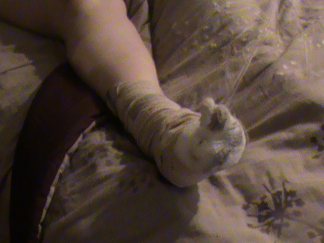
A week ago I had my 4th toe right foot amputated.  It’s still wrapped up.  I have a little phantom pain-but not too much and I think it’s just the nerve remembering-soon to forget.  I had it done in the hospital in the early afternoon and was home by 4pm!
I’ll take a pic of what my foot looks like as soon as I have the bandage wrap taken off.  Here is my right foot-bandage on.
4th toe right foot amputation for neuroma
A little bit about Neuroma’s from feetmd.com:  4th toe right foot amputation for neuroma
amputated toe
amputated toe
julietoetwo julietoethree julietoefour

Morton’s Neuroma

In the foot, there are the long bones (metatarsals) and thin nerves running between them. The nerves split in a Y-shape when they reach the toes. If the metatarsals move abnormally, they can pinch the nerve between them, which causes inflammation and, eventually, permanent nerve damage. Morton’s neuroma is the most common of this type and affects the nerve between the third and fourth toes.

Click here to read more about Morton’s Neuroma»

Other Neuromas

A neuroma is a painful swelling of a nerve, usually in the ball or heel of the foot. Neuromas may occur after a nerve has been injured, either from a traumatic wound or from damage suffered during surgery. Symptoms include sporadic pain; burning, tingling or numbness of one or more toes; and a popping sensation when walking. Pain is often soothed by taking weight off the foot or by massaging the area.

Tarsal Tunnel Syndrome

Tarsal tunnel syndrome is a condition classified by chronic pain in the ankle, foot and toes caused by abnormal pressure on nerve roots. It is similar to carpal tunnel syndrome in the wrist and hand, but is not as common. The specific cause of tarsal tunnel syndrome is not known, but it can be a result of inflamed tissues around the tibial nerve, injury or other conditions that may affect the area.

The main symptom of tarsal tunnel syndrome is tingling or burning pain while standing or walking that starts in the ankle and spreads to the toes. Pain is usually relieved during rest. Doctors diagnose this condition by trying to induce the tingling sensation when tapping the nerve.
Treatment for tarsal tunnel syndrome depends on the cause of the pain but can include anti-inflammatory medication, orthotics, corticosteroid injections or surgery. Surgery is usually used as a last resort to relieve pressure on the nerve.

I had a failed median neurectomy July 12th and this amputation was a last resort for intractable nerve pain.  I’m 52, married 23 years and perfectly willing to take the risk of surgery if it means less pain.  Having several painful conditions, lupus, spondyloarthritis, recurring sacroillitis, osteoarthritis, PN, carpal, tarpal and ulnar neuropathy-this toe was the camel that broke the camel’s back.  That is why I decided on the total solution.  Also between surgeries I have to go off my lupus meds-and THAT is very difficult.  I have experienced a low grade fever almost every day, tons of mouth and nose sores, fatigue, muscle and joint pain, worsening anemia, etc.  This was my third surgery this year.  I am very positive I made a good choice FOR ME.  :)

PART 2:  I WON AT MY SSDI APPEAL HEARING!
After just over three years I had my day in court for social security disability.  I WON my  case.  I had a wonderful lawyer who did a tip-top job of organizing and preparing my medical files which were over two feet high, lol!  I’m still in shock!  I will talk more about this in another post where I hope I can be helpful for other people like me with lupus who are applying for disability.

Here’s the short list:  One thing is to keep all your medical records.  Have a good repore with your doctors, especially your rheumatologist and GP, neurologist if they are a big part of your case, etc.  Prepare them that you are coming to the end of your working days-and hopefully get their support.  Enough to have them fill out paperwork for your lawyer or to write you a letter describing your inability to work and why.

If you worked and had to stop-get a letter from your previous employers.  Hopefully this letter will reflect that due to no fault of your own you had to stop working-that your condition symptoms, doctor appointments and medication side effects all effected your ability to work but that while you did work you were an exemplory employee!  This is very beneficial to your case.
Of course, find a lawyer who specializes in social security disability, hopefully one that has been doing this type of work for many years-a local attorney is always better because they have dealt with the particular judges that you will be seeing should your case go to a hearing.  Any lawyer you do find should work on contingency-with about a 25 to 33% of your backpay reward as a fee.  There is a cap at $6000, so no worries there.  If a lawyer wants to charge you upfront I would look around for another.

PART 3-MY FAVORITE PAIN DOC HIT A VEIN BY ACCIDENT WITH LIDOCAINE DURING A PROCEDURE!  BIG OOPS!

I was considered unresponsive!  First let me say that I adore this doctor-he has given me back quality of life with radiofrequency ablations, cortisone and alcohol shots, and nerve blocks in addition to effective pain management medications.

I have all procedures in office and without sedation.  I’m good about that.  My theory is if your there for dealing with pain, you can make it through the procedures without being knocked out.  That said, this accident KNOCKED ME OUT COLD!

I woke up in a recovery chair confused and bewildered to say the least.  One of the nurses handed me my purse and coffee cup, which I dumped and spilled all over my lap in a dazed state.  They slid the thingy off my finger and said I was done and to go up front and wait for my scripts.  Huh?  I was really out of it.  I knew I was asleep but not why..really strange feeling.

As I wobbled down the hallway I looked back and my doctor half jogged towards me.  He said he accidentally got a blood vessel with the lidocaine.  I had no idea what that meant but it didn’t sound good.  I asked the one question that mattered.  I said, “AM I OK?”   He said “YES”, so I continued my wobble down the hall to check out and make my next appointment.  Really dazed…

Long story short when I got home I noticed I had bandages around my wrist and that both arms were black and blue.  My right wrist was black and blue-looked a little like dirt, and my left wrist where the bandage had been was bruised also but with puncture marks both on the wrist and then up by the inside of my elbows.

My husband called the docs office after this fiasco to find out exactly what happened.  They told him (they read it off the docs report)  that I was unresponsive and that he accidentally injected the lidocaine into a vein.  I still don’t know what that means exactly.  I know I was unconscious since the last thing I heard was “Julie, your doing great-almost done” and then I WOKE UP in a chair.  Huh?  Was I unresponsive other than unconscious?  Did my heart stop?  Did my breathing stop?  And WHAT DID THEY INJECT that they made all these holes in me?

Why did they not explain to me when I woke up, etc.  I was expecting a call back from my doc but he had a nurse call.  I wasn’t happy about that.  She tried to make it sound like it was all normal and no big deal.  SHE wasn’t the one UNRESPONSIVE.  I told her I want to know what happened exactly.  She spent her time defending why the nurses around me when I woke up didn’t know any better since they deal with people who are sedated waking up all the time.  I recanted with the fact THAT I WAS NOT SEDATED EVER DURING A PROCEDURE, so it certainly wasn’t normal for me!!!!!!!
Here is a pic of my wrists when I got home:   DSC00510 DSC00514 DSC00515 DSC00516 DSC00517
Thanks for listening!  I read that lidocaine injections accidentally in veins can cause strokes, arrested breathing, severe hypertension, heart attacks, YIKES!

About Lidocaine Accidental Intravascular Overdose from epilepsy.com:
Lidocaine hydrochloride has proconvulsant and anticonvulsant effects, with CNS effects related to blood concentrations. Low doses (2–3 mg/kg) can terminate status epilepticus.

With increasing blood levels, CNS symptoms and signs of toxicity occur, from perioral numbness, lightheadedness, dizziness, tinnitus, and fine tremors to generalized seizures and coma. In animals, lidocaine produces epileptiform activity that is limited to the amygdala and hippocampus.86
Lidocaine doses that are commonly used for local anesthesia can cause CNS toxicity if they are inadvertently administered intravenously. For example, when administering epidural anesthesia, total doses of 5–8 mg/kg are commonly injected into the epidural space.84 Accidental intravascular injection of this dose can cause epileptic seizures.

In addition to direct intravascular injection and immediate toxicity, systemic lidocaine levels can rise to toxic levels by rapid systemic absorption from the area of injection. This can occur 10 to 20 minutes after injection. Anesthesiologists often add 5 mg/mL of epinephrine to the local anesthetic to decrease systemic absorption and peak serum lidocaine levels. When a regional anesthesia block is successful, early reinjection of local anesthesia can cause toxicity (including seizures), because peak absorption of the first injection occurs while additional medication is injected.
Efforts should be made to deliver minimum amounts of lidocaine to the lower respiratory tract in airway anesthesia (e.g., for bronchoscopy), because its pharmacokinetics at that site are similar to those with intravenous administration.87

High doses of lidocaine cause sedation. Increasing the arterial partial pressure of carbon dioxide (PaCO2) decreases the dose of lidocaine needed to produce a generalized electrical seizure.88 Higher PaCO2 levels increase cerebral blood flow, thus increasing the amount of anesthetic reaching the brain, and may directly excite the amygdala. In contrast to the usual pattern, in which hyperventilation activates seizure activity, hyperventilation may prevent seizures from occurring in patients with lidocaine overdose by decreasing cerebral blood flow.

Lidocaine is injected intravenously to provide local anesthesia (intravenous regional anesthesia or Bier blocks). In this technique, after an extremity is exsanguinated, and the blood supply is arrested by a tourniquet, lidocaine is injected into a vein to provide anesthesia. Doses of lidocaine up to 3 mg/kg of 0.5% solution without preservatives or epinephrine are used. Premature tourniquet release (less than 20 minutes) can result in high systemic lidocaine levels and possible seizure activity. Release after 20 minutes can also be associated with toxicity. Some physicians cycle the deflation of the tourniquet with an intermittent inflation-deflation-inflation cycle in an attempt to decrease rapid absorption of lidocaine from the extremity.

Seizures induced by lidocaine can be terminated with barbiturates.
Etidocaine hydrochloride, a long-acting derivative of lidocaine, as well as mepivacaine and prilocaine hydrochloride, share common pharmacologic properties with lidocaine hydrochloride.
Adapted from: Najjar S, Devinsky O, Rosenberg AD, et al. Procedures in epilepsy patients. In: Ettinger AB and Devinsky O, eds. Managing epilepsy and co-existing disorders. Boston: Butterworth-Heinemann; 2002;499–513. With permission from Elsevier (www.elsevier.com).
Reviewed and revised April 2004 by Steven C. Schachter, MD, epilepsy.com Editorial Board.
This conversation is from ExpertLaw.com about the exact subject of a lidocaine overdose by vein during a pain procedure:
  1. Lidocaine Reaction

    Yesterday I received lidocaine as a local anesthetic while getting a steriod epidural for bulging disc problems in lower spine. During procedure I began to have a severe allergic reaction that required I be taken by ambulance from the clinic to the ER. I was released after 4 hours of observation (and benedryl), but still not feeling very well today.
    The ER doc (not me) raised the possibility of an overdose, but said he asked the doctor who adminstered the lidocaine who said I got the right dose. I suppose there is also the chance the lidocaine was injected directly into a vein by mistake.
    How do I figure out what happened and if there is any fault here? I’m clearly not allergic to lidocaine or the steriod at normal levels since I’ve received both many times before.
  2. Default Re: Lidocaine Reaction

    What kind of severe allergic reaction? Cardiac or itching/swelling?
    You could always get a copy of your medical record since the procedure, your reaction, and the fact that they had to call an ambulance should be documented.
  3. Default Re: Lidocaine Reaction

    Thank you for your response.
    The reaction began on the table as really bad hot flash and nausea after the lidocaine injection. The doctor told me to hold on while he finished the ESI. It got worse when I got up.
    After moving to a chair post-procedure, symptoms of strong dull pain in chest/upper body and difficulty breathing began, and continued to get worse. Doctor initially said this was “normal” and would go away in 10 minutes or so. My BP was high and O2 level was low (based on the finger clamp thing?). When it didn’t go away, I was moved to a laying position, and after another 10 minutes, the uncontrollable, severe shaking started. I think that’s when they called an ambulance and took me to the hospital.
    In the ambulance they gave me a IV dose of benedryl. I also had tingling/itchy hands and blotchy skin.
    After getting to hospital, shaking and chest pain stopped and breathing seemed to return to normal.
    Also, isn’t the fact that they are billing my insurance company for the ambulance documentation enough that I needed that?
  4. Default Re: Lidocaine Reaction

    First, the informed consent you signed before the procedure listed the side effects/risks of the epidural, as well as the medications given during the procedure.
    The fact that you did not have a previous reaction to Lidocaine doesn’t mean you’ll never have a reaction to it. It also does not mean the reaction you had was necessarily caused by Lidocaine.
    The ambulance company billing your insurance is not he kind of documentation you need. You’ll need a copy of the office notes from the procedure, which should contain notation of your reaction and the fact that 911 was necessary. You’ll also need a copy of the hospital ER record.
  5. Default Re: Lidocaine Reaction

    Thanks. I guess one final question, and I don’t mean this to sound cavalier. If I signed the informed consent assuming all responsibility and risks, why do I “need” a copy of anything at this point?
  6. Default Re: Lidocaine Reaction

    You originally asked:
    How do I figure out what happened and if there is any fault here?
    The answer may be in your medical record.
    Thanks. I guess one final question, and I don’t mean this to sound cavalier. If I signed the informed consent assuming all responsibility and risks, why do I “need” a copy of anything at this point?
  7. you don’t assume all responsibility nor waive all rights to seek redress. You signed consent based on known risks and complications. That doesn’t mean if a doctor is negligent you have no redress available (not suggesting there were in this case, simply as explanation of what would not be waived by your consent form)
    THANKS FOR LISTENING EVERYONE!   ((((((((HUGS))))))))                Julie

Thursday, August 8, 2013

Reactive Arthritis-WHAT? “React THIS-Arthritis”!

For me it took 3 bouts of sacroillitis (one was VERY bad-oy vay- couldn't walk, sleep, lay) that comes accompanied with it's pal uveitis and pinkeye. 

My rheumatologist calls with the results of my genetic testing-the gene HLA-B27 locked up the dx after the MRI of lower lumbar and si joint showed inflammation only and no bone fusing-had there been bone fusing a diagnosis of anklylosing spondylitis would have been appropriate.  Thank goodness it wasn't that-well isn't so far-  My grandmother had AS and she was in great pain and hunched over.  She also had undiagnosed lupus, which attacked her nerves, blood and skin the most-just like ME. 

The pain radiating on one side above my butt and under lower back-now known as the si joint is warm to the touch, swollen looking and varies in intensity when it acts up and is painful.  At it's worst I would not hesitate to clobber ya for getting in touching distance, lol.  Sometimes it's one side, sometimes its both and last December, the worst flare I ever had with it, it was both, it also included the pelvic area and I could not MOVE without pain.  Its horrendous.  It comes with uveitis and/or pinkeye.  And what the two have to do with each other, BEATS ME!  But here's the idea the doctors have:

Fast facts

  • Reactive arthritis can affect the heels, toes, fingers, low back, and joints, especially of the knees or ankles.
  • The infection that causes reactive arthritis usually presents (shows up) as diarrhea or as a sexually transmitted disease. But, it can have no symptoms (called asymptomatic).
  • Though it often goes away on its own, reactive arthritis can be prolonged and severe enough to require seeing a specialist.
My eyes with uveitis and scleritis.  Not fun.

What is reactive arthritis?

Reactive arthritis is a painful form of inflammatory arthritis (joint disease due to inflammation). It occurs in reaction to an infection by certain bacteria. Most often, these bacteria are in the genitals (Chlamydia trachomatis) or the bowel (Campylobacter, Salmonella, Shigella and Yersinia). Chlamydia most often transmits by sex. It often has no symptoms, but can cause a pus-like or watery discharge from the genitals. The bowel bacteria can cause diarrhea.
Reactive arthritis can have any or all of these features:
  • Pain and swelling of certain joints, often the knees and/or ankles
  • Swelling and pain at the heels
  • Extensive swelling of the toes or fingers
  • Persistent low back pain, which tends to be worse at night or in the morning
Some patients with this type of arthritis also have eye redness and irritation. Still other signs and symptoms include burning with urination and a rash on the palms or the soles of the feet.

What causes reactive arthritis?

The bacteria induce (cause) arthritis by distorting your body's defense against infections, as well as your genetic environment.
How exactly each of these factors plays a role in the disease likely varies from patient to patient. This is a focus of research.

Who gets reactive arthritis?

The bacteria that cause reactive arthritis are very common. In theory, anyone who becomes infected with these germs might develop reactive arthritis. Yet very few people with bacterial diarrhea actually go on to have serious reactive arthritis.
Musculoskeletal
Signs and symptoms that affect your bones and muscles may include:
  • Joint pain, usually in your knees, ankles and feet
  • Heel pain
  • Pain and swelling at the back of your ankle
  • Swollen toes or fingers, which may look like sausages
  • Pain in your low back or buttocks
Reproductive and urinary
Possible signs and symptoms of your reproductive and urinary systems include:
  • Pain or burning during urination
  • Increased frequency of urination
  • Inflammation of the prostate gland (prostatitis)
  • Inflammation of the cervix (cervicitis)
Eyes, mouth and skin
Signs and symptoms that affect your eyes, mouth and skin may include:
  • Eye inflammation (conjunctivitis)
  • Inflammation of your inner eye (uveitis)
  • Mouth ulcers
  • Skin rashes
Reactive arthritis develops in reaction to an infection in another part of your body, often in your intestines, genitals or urinary tract. You may not be aware of the triggering infection because it may cause only mild symptoms or none at all.
Numerous bacteria can cause reactive arthritis. The most common ones include:
  • Chlamydia
  • Salmonella
  • Shigella
  • Yersinia
  • Campylobacter
Reactive arthritis isn't contagious. However, the bacteria that cause it can be transmitted sexually or in contaminated food. But only a few of the people who are exposed to these bacteria develop reactive arthritis.
Also called Reiter's Syndrome-here is more info:
https://www.mayoclinic.com/health/reactive-arthritis/DS00486
http://www.rheumatology.org/Practice/Clinical/Patients/Diseases_And_Conditions/Reactive_Arthritis/


Tuesday, July 23, 2013

for·ti·tude

/ˈfôrtiˌto͞od/

Noun
Courage in pain or adversity: “she endured her illness with great fortitude”.

I was writing an email to my father and out popped the word “fortitude”.  “Wish I had the fortitude” I wrote.  I know what it means but I looked it up anyway. Its definition pops out AT ME.  (see above)  And I can’t help thinking it’s not a coincidence.  I’m not enduring my illness with any for·ti·tude  at all and I should be.  I am more of an unorganized mess-lately hit with that dreaded brain fog forgetting what I’m doing and what I’m saying, names, places, faces, details.  Not enough of a help to my family.  I need to step up higher.  My husband is tired.  My son needs my help.  

Disability hearing is coming up in September.  The board of education kicked back my dismissed Sallie Mae school loans because my GP is a DO.  Go figure.  Alot going on and alot coming up.  My foot surgery seems failed and I will likely need two more, one to kill the other side of the nerve and one to take the toe.  The familiar pain has returned along with my unsettledness & has run off with my for·ti·tude.
Tomorrow is another day.  Tally Ho!