Friday, October 25, 2013

The Nerve of THIS NERVE-Failed Amputation-Going for Round 3!

The #NERVE of THIS NERVE; #Amputation Failed! Not giving up!
Yesterday I saw my surgeon-the amputation was a fail. The darn median nerve continues to curse my days and sleep. It went five days nonstop. Doc gave me no less than EIGHT lidocaine shots yesterday to shut it down. By the time he was done I couldn't feel any part of my right foot EXCEPT THE NERVE PAIN. I mean, it practically defies nature. The other possibility according to the million dollar nerve conduction equipment is that there are severely damaged small to medium fiber peripheral nerves in the skin that was used to close up the amputation.

Anyway, THIS IS WAR (as my BIL used to say)

End of story is I'm going in for another surgery. I'll let you know when it is. He's cutting the median nerve in two new places and ripping out the skin used to cover up where the toe is amputated and stretching what's left to use. He may have to graft later if it doesn't take and I get a wound problem. Ah, another risk I'm willing to take-wound problems. Like I said, "THIS IS WAR" and I'm not backin down until this nerve is gone for good.

I know what your thinking but I'm actually very lucky to have a surgeon willing to go thrice trying to help me. This is his last suggestion, after that-he is outta ideas. You've got to remember I am a complicated case with circulating immune complexes causing inflammation in my blood vessels that press on nerves and damage them. This is my particular present from lupus.
So.......that's whats happening. :)

Tuesday, September 17, 2013

Toe Amputated-Goodbye Neuroma! Hello Successful SSDI Hearing! Goodbye Lidocaine Overdose!


So much has happened! I’ll break it up in Parts.  Part 1-toe amputation.  Part 2- I WON at my disability appeal hearing for SSDI.  Part 3-My favorite pain doc accidentally hit my vein with lidocaine and I became UNRESPONSIVE! 
 
PART 1 – TOE AMPUTATION
A week ago I had my 4th toe right foot amputated.  It’s still wrapped up.  I have a little phantom pain-but not too much and I think it’s just the nerve remembering-soon to forget.  I had it done in the hospital in the early afternoon and was home by 4pm!
I’ll take a pic of what my foot looks like as soon as I have the bandage wrap taken off.  Here is my right foot-bandage on.
4th toe right foot amputation for neuroma
A little bit about Neuroma’s from feetmd.com:  4th toe right foot amputation for neuroma
amputated toe
amputated toe
julietoetwo julietoethree julietoefour

Morton’s Neuroma

In the foot, there are the long bones (metatarsals) and thin nerves running between them. The nerves split in a Y-shape when they reach the toes. If the metatarsals move abnormally, they can pinch the nerve between them, which causes inflammation and, eventually, permanent nerve damage. Morton’s neuroma is the most common of this type and affects the nerve between the third and fourth toes.

Click here to read more about Morton’s Neuroma»

Other Neuromas

A neuroma is a painful swelling of a nerve, usually in the ball or heel of the foot. Neuromas may occur after a nerve has been injured, either from a traumatic wound or from damage suffered during surgery. Symptoms include sporadic pain; burning, tingling or numbness of one or more toes; and a popping sensation when walking. Pain is often soothed by taking weight off the foot or by massaging the area.

Tarsal Tunnel Syndrome

Tarsal tunnel syndrome is a condition classified by chronic pain in the ankle, foot and toes caused by abnormal pressure on nerve roots. It is similar to carpal tunnel syndrome in the wrist and hand, but is not as common. The specific cause of tarsal tunnel syndrome is not known, but it can be a result of inflamed tissues around the tibial nerve, injury or other conditions that may affect the area.

The main symptom of tarsal tunnel syndrome is tingling or burning pain while standing or walking that starts in the ankle and spreads to the toes. Pain is usually relieved during rest. Doctors diagnose this condition by trying to induce the tingling sensation when tapping the nerve.
Treatment for tarsal tunnel syndrome depends on the cause of the pain but can include anti-inflammatory medication, orthotics, corticosteroid injections or surgery. Surgery is usually used as a last resort to relieve pressure on the nerve.

I had a failed median neurectomy July 12th and this amputation was a last resort for intractable nerve pain.  I’m 52, married 23 years and perfectly willing to take the risk of surgery if it means less pain.  Having several painful conditions, lupus, spondyloarthritis, recurring sacroillitis, osteoarthritis, PN, carpal, tarpal and ulnar neuropathy-this toe was the camel that broke the camel’s back.  That is why I decided on the total solution.  Also between surgeries I have to go off my lupus meds-and THAT is very difficult.  I have experienced a low grade fever almost every day, tons of mouth and nose sores, fatigue, muscle and joint pain, worsening anemia, etc.  This was my third surgery this year.  I am very positive I made a good choice FOR ME.  :)

PART 2:  I WON AT MY SSDI APPEAL HEARING!
After just over three years I had my day in court for social security disability.  I WON my  case.  I had a wonderful lawyer who did a tip-top job of organizing and preparing my medical files which were over two feet high, lol!  I’m still in shock!  I will talk more about this in another post where I hope I can be helpful for other people like me with lupus who are applying for disability.

Here’s the short list:  One thing is to keep all your medical records.  Have a good repore with your doctors, especially your rheumatologist and GP, neurologist if they are a big part of your case, etc.  Prepare them that you are coming to the end of your working days-and hopefully get their support.  Enough to have them fill out paperwork for your lawyer or to write you a letter describing your inability to work and why.

If you worked and had to stop-get a letter from your previous employers.  Hopefully this letter will reflect that due to no fault of your own you had to stop working-that your condition symptoms, doctor appointments and medication side effects all effected your ability to work but that while you did work you were an exemplory employee!  This is very beneficial to your case.
Of course, find a lawyer who specializes in social security disability, hopefully one that has been doing this type of work for many years-a local attorney is always better because they have dealt with the particular judges that you will be seeing should your case go to a hearing.  Any lawyer you do find should work on contingency-with about a 25 to 33% of your backpay reward as a fee.  There is a cap at $6000, so no worries there.  If a lawyer wants to charge you upfront I would look around for another.

PART 3-MY FAVORITE PAIN DOC HIT A VEIN BY ACCIDENT WITH LIDOCAINE DURING A PROCEDURE!  BIG OOPS!

I was considered unresponsive!  First let me say that I adore this doctor-he has given me back quality of life with radiofrequency ablations, cortisone and alcohol shots, and nerve blocks in addition to effective pain management medications.

I have all procedures in office and without sedation.  I’m good about that.  My theory is if your there for dealing with pain, you can make it through the procedures without being knocked out.  That said, this accident KNOCKED ME OUT COLD!

I woke up in a recovery chair confused and bewildered to say the least.  One of the nurses handed me my purse and coffee cup, which I dumped and spilled all over my lap in a dazed state.  They slid the thingy off my finger and said I was done and to go up front and wait for my scripts.  Huh?  I was really out of it.  I knew I was asleep but not why..really strange feeling.

As I wobbled down the hallway I looked back and my doctor half jogged towards me.  He said he accidentally got a blood vessel with the lidocaine.  I had no idea what that meant but it didn’t sound good.  I asked the one question that mattered.  I said, “AM I OK?”   He said “YES”, so I continued my wobble down the hall to check out and make my next appointment.  Really dazed…

Long story short when I got home I noticed I had bandages around my wrist and that both arms were black and blue.  My right wrist was black and blue-looked a little like dirt, and my left wrist where the bandage had been was bruised also but with puncture marks both on the wrist and then up by the inside of my elbows.

My husband called the docs office after this fiasco to find out exactly what happened.  They told him (they read it off the docs report)  that I was unresponsive and that he accidentally injected the lidocaine into a vein.  I still don’t know what that means exactly.  I know I was unconscious since the last thing I heard was “Julie, your doing great-almost done” and then I WOKE UP in a chair.  Huh?  Was I unresponsive other than unconscious?  Did my heart stop?  Did my breathing stop?  And WHAT DID THEY INJECT that they made all these holes in me?

Why did they not explain to me when I woke up, etc.  I was expecting a call back from my doc but he had a nurse call.  I wasn’t happy about that.  She tried to make it sound like it was all normal and no big deal.  SHE wasn’t the one UNRESPONSIVE.  I told her I want to know what happened exactly.  She spent her time defending why the nurses around me when I woke up didn’t know any better since they deal with people who are sedated waking up all the time.  I recanted with the fact THAT I WAS NOT SEDATED EVER DURING A PROCEDURE, so it certainly wasn’t normal for me!!!!!!!
Here is a pic of my wrists when I got home:   DSC00510 DSC00514 DSC00515 DSC00516 DSC00517
Thanks for listening!  I read that lidocaine injections accidentally in veins can cause strokes, arrested breathing, severe hypertension, heart attacks, YIKES!

About Lidocaine Accidental Intravascular Overdose from epilepsy.com:
Lidocaine hydrochloride has proconvulsant and anticonvulsant effects, with CNS effects related to blood concentrations. Low doses (2–3 mg/kg) can terminate status epilepticus.

With increasing blood levels, CNS symptoms and signs of toxicity occur, from perioral numbness, lightheadedness, dizziness, tinnitus, and fine tremors to generalized seizures and coma. In animals, lidocaine produces epileptiform activity that is limited to the amygdala and hippocampus.86
Lidocaine doses that are commonly used for local anesthesia can cause CNS toxicity if they are inadvertently administered intravenously. For example, when administering epidural anesthesia, total doses of 5–8 mg/kg are commonly injected into the epidural space.84 Accidental intravascular injection of this dose can cause epileptic seizures.

In addition to direct intravascular injection and immediate toxicity, systemic lidocaine levels can rise to toxic levels by rapid systemic absorption from the area of injection. This can occur 10 to 20 minutes after injection. Anesthesiologists often add 5 mg/mL of epinephrine to the local anesthetic to decrease systemic absorption and peak serum lidocaine levels. When a regional anesthesia block is successful, early reinjection of local anesthesia can cause toxicity (including seizures), because peak absorption of the first injection occurs while additional medication is injected.
Efforts should be made to deliver minimum amounts of lidocaine to the lower respiratory tract in airway anesthesia (e.g., for bronchoscopy), because its pharmacokinetics at that site are similar to those with intravenous administration.87

High doses of lidocaine cause sedation. Increasing the arterial partial pressure of carbon dioxide (PaCO2) decreases the dose of lidocaine needed to produce a generalized electrical seizure.88 Higher PaCO2 levels increase cerebral blood flow, thus increasing the amount of anesthetic reaching the brain, and may directly excite the amygdala. In contrast to the usual pattern, in which hyperventilation activates seizure activity, hyperventilation may prevent seizures from occurring in patients with lidocaine overdose by decreasing cerebral blood flow.

Lidocaine is injected intravenously to provide local anesthesia (intravenous regional anesthesia or Bier blocks). In this technique, after an extremity is exsanguinated, and the blood supply is arrested by a tourniquet, lidocaine is injected into a vein to provide anesthesia. Doses of lidocaine up to 3 mg/kg of 0.5% solution without preservatives or epinephrine are used. Premature tourniquet release (less than 20 minutes) can result in high systemic lidocaine levels and possible seizure activity. Release after 20 minutes can also be associated with toxicity. Some physicians cycle the deflation of the tourniquet with an intermittent inflation-deflation-inflation cycle in an attempt to decrease rapid absorption of lidocaine from the extremity.

Seizures induced by lidocaine can be terminated with barbiturates.
Etidocaine hydrochloride, a long-acting derivative of lidocaine, as well as mepivacaine and prilocaine hydrochloride, share common pharmacologic properties with lidocaine hydrochloride.
Adapted from: Najjar S, Devinsky O, Rosenberg AD, et al. Procedures in epilepsy patients. In: Ettinger AB and Devinsky O, eds. Managing epilepsy and co-existing disorders. Boston: Butterworth-Heinemann; 2002;499–513. With permission from Elsevier (www.elsevier.com).
Reviewed and revised April 2004 by Steven C. Schachter, MD, epilepsy.com Editorial Board.
This conversation is from ExpertLaw.com about the exact subject of a lidocaine overdose by vein during a pain procedure:
  1. Lidocaine Reaction

    Yesterday I received lidocaine as a local anesthetic while getting a steriod epidural for bulging disc problems in lower spine. During procedure I began to have a severe allergic reaction that required I be taken by ambulance from the clinic to the ER. I was released after 4 hours of observation (and benedryl), but still not feeling very well today.
    The ER doc (not me) raised the possibility of an overdose, but said he asked the doctor who adminstered the lidocaine who said I got the right dose. I suppose there is also the chance the lidocaine was injected directly into a vein by mistake.
    How do I figure out what happened and if there is any fault here? I’m clearly not allergic to lidocaine or the steriod at normal levels since I’ve received both many times before.
  2. Default Re: Lidocaine Reaction

    What kind of severe allergic reaction? Cardiac or itching/swelling?
    You could always get a copy of your medical record since the procedure, your reaction, and the fact that they had to call an ambulance should be documented.
  3. Default Re: Lidocaine Reaction

    Thank you for your response.
    The reaction began on the table as really bad hot flash and nausea after the lidocaine injection. The doctor told me to hold on while he finished the ESI. It got worse when I got up.
    After moving to a chair post-procedure, symptoms of strong dull pain in chest/upper body and difficulty breathing began, and continued to get worse. Doctor initially said this was “normal” and would go away in 10 minutes or so. My BP was high and O2 level was low (based on the finger clamp thing?). When it didn’t go away, I was moved to a laying position, and after another 10 minutes, the uncontrollable, severe shaking started. I think that’s when they called an ambulance and took me to the hospital.
    In the ambulance they gave me a IV dose of benedryl. I also had tingling/itchy hands and blotchy skin.
    After getting to hospital, shaking and chest pain stopped and breathing seemed to return to normal.
    Also, isn’t the fact that they are billing my insurance company for the ambulance documentation enough that I needed that?
  4. Default Re: Lidocaine Reaction

    First, the informed consent you signed before the procedure listed the side effects/risks of the epidural, as well as the medications given during the procedure.
    The fact that you did not have a previous reaction to Lidocaine doesn’t mean you’ll never have a reaction to it. It also does not mean the reaction you had was necessarily caused by Lidocaine.
    The ambulance company billing your insurance is not he kind of documentation you need. You’ll need a copy of the office notes from the procedure, which should contain notation of your reaction and the fact that 911 was necessary. You’ll also need a copy of the hospital ER record.
  5. Default Re: Lidocaine Reaction

    Thanks. I guess one final question, and I don’t mean this to sound cavalier. If I signed the informed consent assuming all responsibility and risks, why do I “need” a copy of anything at this point?
  6. Default Re: Lidocaine Reaction

    You originally asked:
    How do I figure out what happened and if there is any fault here?
    The answer may be in your medical record.
    Thanks. I guess one final question, and I don’t mean this to sound cavalier. If I signed the informed consent assuming all responsibility and risks, why do I “need” a copy of anything at this point?
  7. you don’t assume all responsibility nor waive all rights to seek redress. You signed consent based on known risks and complications. That doesn’t mean if a doctor is negligent you have no redress available (not suggesting there were in this case, simply as explanation of what would not be waived by your consent form)
    THANKS FOR LISTENING EVERYONE!   ((((((((HUGS))))))))                Julie

Thursday, August 8, 2013

Reactive Arthritis-WHAT? “React THIS-Arthritis”!

For me it took 3 bouts of sacroillitis (one was VERY bad-oy vay- couldn't walk, sleep, lay) that comes accompanied with it's pal uveitis and pinkeye. 

My rheumatologist calls with the results of my genetic testing-the gene HLA-B27 locked up the dx after the MRI of lower lumbar and si joint showed inflammation only and no bone fusing-had there been bone fusing a diagnosis of anklylosing spondylitis would have been appropriate.  Thank goodness it wasn't that-well isn't so far-  My grandmother had AS and she was in great pain and hunched over.  She also had undiagnosed lupus, which attacked her nerves, blood and skin the most-just like ME. 

The pain radiating on one side above my butt and under lower back-now known as the si joint is warm to the touch, swollen looking and varies in intensity when it acts up and is painful.  At it's worst I would not hesitate to clobber ya for getting in touching distance, lol.  Sometimes it's one side, sometimes its both and last December, the worst flare I ever had with it, it was both, it also included the pelvic area and I could not MOVE without pain.  Its horrendous.  It comes with uveitis and/or pinkeye.  And what the two have to do with each other, BEATS ME!  But here's the idea the doctors have:

Fast facts

  • Reactive arthritis can affect the heels, toes, fingers, low back, and joints, especially of the knees or ankles.
  • The infection that causes reactive arthritis usually presents (shows up) as diarrhea or as a sexually transmitted disease. But, it can have no symptoms (called asymptomatic).
  • Though it often goes away on its own, reactive arthritis can be prolonged and severe enough to require seeing a specialist.
My eyes with uveitis and scleritis.  Not fun.

What is reactive arthritis?

Reactive arthritis is a painful form of inflammatory arthritis (joint disease due to inflammation). It occurs in reaction to an infection by certain bacteria. Most often, these bacteria are in the genitals (Chlamydia trachomatis) or the bowel (Campylobacter, Salmonella, Shigella and Yersinia). Chlamydia most often transmits by sex. It often has no symptoms, but can cause a pus-like or watery discharge from the genitals. The bowel bacteria can cause diarrhea.
Reactive arthritis can have any or all of these features:
  • Pain and swelling of certain joints, often the knees and/or ankles
  • Swelling and pain at the heels
  • Extensive swelling of the toes or fingers
  • Persistent low back pain, which tends to be worse at night or in the morning
Some patients with this type of arthritis also have eye redness and irritation. Still other signs and symptoms include burning with urination and a rash on the palms or the soles of the feet.

What causes reactive arthritis?

The bacteria induce (cause) arthritis by distorting your body's defense against infections, as well as your genetic environment.
How exactly each of these factors plays a role in the disease likely varies from patient to patient. This is a focus of research.

Who gets reactive arthritis?

The bacteria that cause reactive arthritis are very common. In theory, anyone who becomes infected with these germs might develop reactive arthritis. Yet very few people with bacterial diarrhea actually go on to have serious reactive arthritis.
Musculoskeletal
Signs and symptoms that affect your bones and muscles may include:
  • Joint pain, usually in your knees, ankles and feet
  • Heel pain
  • Pain and swelling at the back of your ankle
  • Swollen toes or fingers, which may look like sausages
  • Pain in your low back or buttocks
Reproductive and urinary
Possible signs and symptoms of your reproductive and urinary systems include:
  • Pain or burning during urination
  • Increased frequency of urination
  • Inflammation of the prostate gland (prostatitis)
  • Inflammation of the cervix (cervicitis)
Eyes, mouth and skin
Signs and symptoms that affect your eyes, mouth and skin may include:
  • Eye inflammation (conjunctivitis)
  • Inflammation of your inner eye (uveitis)
  • Mouth ulcers
  • Skin rashes
Reactive arthritis develops in reaction to an infection in another part of your body, often in your intestines, genitals or urinary tract. You may not be aware of the triggering infection because it may cause only mild symptoms or none at all.
Numerous bacteria can cause reactive arthritis. The most common ones include:
  • Chlamydia
  • Salmonella
  • Shigella
  • Yersinia
  • Campylobacter
Reactive arthritis isn't contagious. However, the bacteria that cause it can be transmitted sexually or in contaminated food. But only a few of the people who are exposed to these bacteria develop reactive arthritis.
Also called Reiter's Syndrome-here is more info:
https://www.mayoclinic.com/health/reactive-arthritis/DS00486
http://www.rheumatology.org/Practice/Clinical/Patients/Diseases_And_Conditions/Reactive_Arthritis/


Tuesday, July 23, 2013

for·ti·tude

/ˈfôrtiˌto͞od/

Noun
Courage in pain or adversity: “she endured her illness with great fortitude”.

I was writing an email to my father and out popped the word “fortitude”.  “Wish I had the fortitude” I wrote.  I know what it means but I looked it up anyway. Its definition pops out AT ME.  (see above)  And I can’t help thinking it’s not a coincidence.  I’m not enduring my illness with any for·ti·tude  at all and I should be.  I am more of an unorganized mess-lately hit with that dreaded brain fog forgetting what I’m doing and what I’m saying, names, places, faces, details.  Not enough of a help to my family.  I need to step up higher.  My husband is tired.  My son needs my help.  

Disability hearing is coming up in September.  The board of education kicked back my dismissed Sallie Mae school loans because my GP is a DO.  Go figure.  Alot going on and alot coming up.  My foot surgery seems failed and I will likely need two more, one to kill the other side of the nerve and one to take the toe.  The familiar pain has returned along with my unsettledness & has run off with my for·ti·tude.
Tomorrow is another day.  Tally Ho!

Sunday, July 21, 2013

The EYES have it! Uveitis, that is!


ps.  Does the inflammation highway ever run out of track?  I don’t think so!  Todays stop boys and girls will be recurring anterior uveitis.  You-vee itis?  Me-vee-itis?  We all you-me-itis!
This is my second round of pretty bad uveitis-bad enough for the men in white to give me an iv of steroids and a shot in the eyes of the same.  YES, I said a Shot In The Eyes.
My last bout-December 2012 came accompanied with sacroiliitis, alias inflammatory arthritis, alias “are you kidding me” kind of pain.  So far it’s starting about the same.  While uveitis can come to the inflammation party on its own-there are alot of conditions that also can cause it.  In my case docs just blamed the L-Word although my grandmother had ankylosing spondylitis-and she may have set a genetic trap for me.  lol.  There’s a genetic marker-it is called HLA-B27 tissue type and testing is appropriate if you have both sacroiliitis and chronic recurring anterior uveitis.
Here’s what to look for in the inflammation of the eye:  uveitispic
And here’s my eyes:  juliejuly 'juliemalarjuly2013

Of course the uveitis highjacked a malar rash and low grade fever  on it’s way to rain on my surgery recovery parade.  Just when I thought it was safe to start dibbling and dabbling with projects and posts.  It’s getting better now by leaps and bounds thanks to the evil but wonderful prednisone and steroid drops.  All the white floaters are gone (I CAN SEE AGAIN) and the pressure and inflammatory pain is down!  Yippeee!  I don’t know about you-but lupus might be the great imitator-however I am the great procrastinator-and that trumps lupus.  ;)
Here is some info about Uveitis (there is more than one kind) and also while playing around I found a new medicine in trial stages for eye inflammation.  This is potentially sight-saving.  And unfortunately-once lupus stops at the door of the eyes, it comes back often to the party.
From Mayo.com:
The signs, symptoms and characteristics of uveitis include:
  • Eye redness
  • Eye pain
  • Light sensitivity
  • Blurred vision
  • Dark, floating spots in your field of vision (floaters)
  • Decreased vision
  • Whitish area (hypopyon) inside the eye in front of the lower part of the colored area of the eye (iris)
The site of uveitis varies and is described by where in the eye it occurs.
  • Anterior uveitis affects the front of your eye (also called iritis).
  • Posterior uveitis affects the back of your eye (also called choroiditis).
  • Intermediary uveitis affects the ciliary body (also called cyclitis).
  • Panuveitis occurs when all layers of the uvea are inflamed.
In any of these conditions, the jelly-like material in the center of your eye (vitreous) can also become inflamed and infiltrated with inflammatory cells.
Symptoms may occur suddenly and get worse quickly, though in some cases, symptoms develop gradually. Symptoms may be noticeable in one or both eyes.
When to see a doctor
Contact your doctor if you think you may have symptoms of uveitis. Your doctor may refer you to an eye specialist (ophthalmologist). If you’re having significant eye pain and new vision problems, seek prompt medical attention.
If uveitis is caused by an underlying condition, treatment will focus on that specific condition. The goal of treatment is to reduce the inflammation in your eye.
Treatment of uveitis may include:
  • Anti-inflammatory medication. Your doctor may prescribe anti-inflammatory medication, such as a corticosteroid, to treat your uveitis. This medication may be given as eyedrops. Or, you may be given corticosteroid pills or an injection into the eye. For people with difficult-to-treat posterior uveitis, a device that’s implanted in your eye may be an option. This device slowly releases corticosteroid medication into your eye for about 2 1/2 years.
  • Antibiotic or antiviral medication. If uveitis is caused by an infection, antibiotics, antiviral medications or other medicines may be given with or without corticosteroids to bring the infection under control.
  • Immunosuppressive or cell-destroying (cytotoxic) medication. Immunosuppressive or cytotoxic agents may be necessary if your uveitis doesn’t respond well to corticosteroids or becomes severe enough to threaten your vision.
  • Surgery. Vitrectomy — surgery to remove some of the jelly-like material in your eye (vitreous) — may be necessary both for diagnosis and management of your uveitis. A small sample of the vitreous can help identify a specific cause of eye inflammation, such as a virus, bacterium or lymphoma. The procedure may also be used to remove developing scar tissue in the vitreous.
The part of your eye affected by uveitis — either the front (anterior) or back (posterior) of the uvea — may determine how quickly your eye heals. Uveitis affecting the back of your eye tends to heal more slowly than uveitis in the front of the eye. Severe inflammation takes longer to clear up than mild inflammation does.
Uveitis can come back. Make an appointment with your doctor if any of your symptoms reappear after successful treatment.

Eye Disease: Preliminary Findings On Uveitis Drug

Aug. 22, 2012 — Shree Kurup, M.D., director of research in the ophthalmology department at Wake Forest Baptist Medical Center, will present the preliminary findings of a Phase 3 clinical trial on a new drug for the treatment of uveitis, a serious inflammatory condition of the uvea, the middle layer of the eye that provides most of the blood supply to the retina.
Link to Clinical Trials for NEW uveitis medication! 

Monday, July 15, 2013

L for LUPUS, N for Neurectomy & No More Pain??

juliehospitalI had what is called a median nerve neurectomy.  My orthopedic surgeon snipped the median nerve in my right foot that leads to the bottom of the fourth toe (on the left side of the toe) with the hopes and intentions of killing the nerve dead!  That toe has certainly been the bane of my existence for the last five years.  Sometimes something so small can turn into something so big and painful.  They call it a Morton's Neuroma.  Likely mine was caused by inflammation in the connective tissue of my feet-plantar fasciitis.  At one time my PF was so bad I couldn't walk.  Cortisone shots right into the plantar gave me back my mobility but the shots into that toe and median nerve stopped working and the zapping and stinging and sharp pains continued. 

I was lucky.  I found an orthopedic surgeon that specializes in FEET.  He went in through the top of the foot and snipped the median nerve right down the middle to the back of the fourth toe right foot.  Here's a better explanation of what a Morton's Neuroma is and a short video animation I found.
Lupus is different in every patient.  Mine likes to attack my peripheral and cranial nerves, skin and blood.  This is just one example of what lupus can do.

From myfootshop.com: 
Morton's Neuroma is a painful condition of the forefoot that is caused by the entrapment of the common intermetatarsal nerve as it passes through the forefoot to the toes. This condition was first described by Dr. Morton, a Viennese physician, in 1876. The most common location for Morton's neuroma is between the third and fourth toes. The second most common location is between the second and third toes.

To help understand this condition a bit better, let's break down the word neuroma into its' root form. Neuro relates to the nervous system and in this case, we are describing a portion of the peripheral nervous system. The suffix 'oma' is the Latin term that defines a tumor or swelling that is of primary origin. Put the terms together and what is described is a tumor or swelling of a peripheral nerve. Interestingly, a Morton's neuroma is not truly a tumor, but more accurately, a nerve entrapment. Although the term neuroma is somewhat inaccurate in describing this condition, the term neuroma is still used today to describe this unique nerve entrapment.  Other terms may be used to describe Morton's neuroma.  These terms include intermetatarsal neuroma, Morton's metatarsalgia, Morton's neuralgia, plantar neuroma, intermetatarsal perineural fibroma or intermetatarsal nerve entrapment. 

What causes Morton's Neuroma? Why does the intermetatarsal nerve become entrapped? Clinicians and surgeons recognize a number of factors that may aggravate Morton's neuroma, but the primary cause of Morton's neuroma remains elusive. Shoes that are tight in the forefoot will contribute to the symptoms of Morton's Neuroma by binding the forefoot and compressing the common intermetatarsal nerve. High heels will also act to increase the ground reactive forces. Ground reactive force is the amount of force generated as the foot pushes against a fixed surface like the floor. With high heels, the amount and focus of ground reactive force increases since weight bearing is focused in a smaller area (just the forefoot). A higher heel also puts the common intermetatarsal nerve under tension, making it more prone to injury. Activities such as squatting will increase the ground reactive force applied to the plantar foot and aggravate the symptoms of Morton's neuroma. And finally, clinician also agree that hypermobility of the forefoot can contribute to the formation of Morton's neuroma.

Treatment of Morton's neuroma
It's interesting to note that until the early 1990's, we treated Morton's neuroma in same way as described by Dr. Morton some 100 years ago. But over the past ten years, our understanding and treatment of neuromas has changed dramatically. Our understanding of Morton's neuroma as an entrapment and not a tumor can be attributed to the work of Steve Barrett, DPM. Dr. Barrett was the first to take a critical look at Morton's neuroma and describe it as an entrapment rather than a tumor.

What Dr. Barrett recognized was that the common intermetatarsal nerve is sometimes prone to becoming entrapped as it passes beneath the intermetatarsal ligament. This was a new concept for us in light of the fact that we had considered Morton's neuroma a tumor. In fact, Dr. Barrett's findings enabled us to recognize Morton's Neuroma to be similar to other nerve entrapments such as carpal tunnel syndrome or tarsal tunnel syndrome. Subsequently, the treatment of Morton's Neuroma has been slowly changing over the last ten years as the result of a new endoscopic surgical procedure first described by Dr. Barrett.

Diagnostic testing to evaluate Morton's neuroma includes plain x-rays, diagnostic ultrasound and MRI. Plain x-rays are not actually used to visualize the nerve, but are use rather to screen for bone and joint pathology adjacent to the nerve. Metatarsal fractures, Freiberg's infraction and osteoarthritis are common conditions that can influence the behavior of Morton's neuroma and need to be evaluated with x-ray.

Several authors have suggested that the efficacy of MRI and ultrasound as diagnostic tools are comparable when evaluating patients for Morton's neuroma. Diagnostic ultrasound is significantly less expensive and much more readily available compared to MRI. Kankanala et. al described a 91.48% pre-op predictive value for diagnostic ultrasound when screening for Morton's neuroma.

Conservative care of Morton's neuroma can be quite successful. 70% or more of new Morton's neuroma patients respond to simple changes in shoes such as a wider toe box. Shoe padding can also help treat Morton's neuroma. Metatarsal pads are an important tool for patients with Morton's neuroma symptoms. A metatarsal pad is a small lift that is positioned in the shoe just proximal (behind) the weight bearing surface of the metatarsal bones. A metatarsal pad lifts and separates the metatarsal bones thereby decreasing the pressure on the intermetatarsal nerve. Some prefabricated arch supports come with a metatarsal pad already seated in the correct position. Using inserts with a metatarsal pad is sometimes the easier way to use a met pad because they can be easily moved from shoe to shoe. Also, by using an insert with a fixed metatarsal pad, the position of the met pad is always in the correct location.

Other non-surgical methods of treating Morton's neuroma include injectable cortisone and chemical sclerosis of the intermetatarsal nerve. Cortisone has been used successfully for years in treating Morton's neuroma. Although the use of cortisone does not actually treat or change the entrapment of the intermetatarsal nerve, cortisone can decrease inflammation and swelling of the nerve, resulting in a decrease in pain. Care should be exercised when using cortisone injections noting that excessive cortisone injections can thin the plantar fat pad of the foot.

Sclerosis of the nerve (also called chemical neuro-ablation or chemical neurolysis) can be performed in the office using a number of different solutions, most commonly dilute (4%) alcohol. Multiple sclerosing injections are used to destroy the contents of the peripheral nerve. A series of injections are employed, each injection separated by a period of 7-10 days. The total number of injections may vary from 3 to 7. The success rates of injectable sclerosing solutions have been reported to be as high as 60-90%. Chemical neurolysis is also a great tool for failed neuroma surgeries where a stump neuroma has formed.

The intent of chemical neurolysis is to destroy the internal contents while preserving the external sheath of the nerve. This would be a bit like removing the copper wire in an electrical wire while preserving the plastic outer insulation or cover of the wire. The reason that this is important is due to the fact that peripheral nerve will regenerate over time. With the nerve sheath intact, regeneration of the nerve is possible in a controlled manner utilizing the existing sheath. By contrast, removal of the nerve by surgery results in the nerve regenerating and the formation of a mass of scar tissue called a stump neuroma. Knowing that peripheral nerve may regenerate also means that sclerosing injections may need to be repeated at some point in the future. The percentage of repeat sclerosing injections varies but is overall quite low.

Another new technique used to treat Morton's neuroma is called cryogenic neuroablation. Cryo surgery is surgery that uses extremely cold instrumentation to selectively destroy tissue. Cryosurgery has been used commonly to destroy superficial skin lesions such as warts and moles.  The technique uses what is referred to as the Joule-Thompson effect. The Joule-Thompson effect occurs when a gas is passed through an area where it may expand. As the gas expands, it cools to approximately -70 degrees centigrade . In the case of cryogenic neuroablation, the expansion of the gas is controlled in a 5.5 mm probe that freezes and subsequently destroys the nerve tissue.

In the cryogenic ablation study carried out by Drs. Caporusso, Fallet and Savoy-Moore, thirty one neuromas were treated in 20 patients. All procedures were performed in an office setting. The procedure used a small amount of local anesthetic to numb the skin to allow the passage of a 12-gauge cannula through the skin. A nerve stimulator was passed through the cannula to locate the nerve. Once the position of the nerve was established, two three minute freeze sessions were utilized to destroy the nerve tissue. A sterile dressing was applied to the site and the patient was dismissed without the need for pain medication. The study cites a 65% success rate.

Dr. Morton's original treatment plan as described in 1876 included changes in shoes, multiple injections of cortisone and if necessary, complete excision of the common intermetatarsal nerve. We've mentioned before that Morton's neuroma is a nerve entrapment much like carpal tunnel. Now let's see if we can apply Dr. Morton's treatment plan to any other nerve entrapment such as carpal tunnel syndrome. Perhaps we'd splint the wrist, try some injectable cortisone, but completely excise the nerve? No way. But that's what's been done for the past 100 years for Morton's Neuroma. Post-op complications were common and included thinning of the plantar fat pad and loss of sensation in the 3rd and 4th toes.

The introduction of Dr. Barrett's EDIN procedure has revolutionized the treatment of Morton's Neuroma and really represents the first unique contribution to treating this condition in over 100 years. The EDIN procedure stands for endoscopic decompression of the common intermetatarsal nerve. Interestingly enough, Steve describes first thinking about this procedure as he watched another surgeon perform an endoscopic carpal tunnel surgery. Steve recognized the problem to be the ligament and not the nerve. The EDIN procedure selectively releases the ligament and leaves the nerve intact.

The EDIN procedure provides us with a new alternative. In the past we knew that the traditional surgery used to treat Morton's Neuroma, called a neurectomy, was destructive and carried with it a number of post-op complications. Therefore, we would tend to use excessive amounts of cortisone to avoid surgery. The EDIN procedure provides a new alternative using non-invasive endoscopic techniques that usually return patients to activities much sooner than the traditional surgery. And, what I find most helpful is the fact that it enables us to use less cortisone, thereby avoiding fat pad atrophy. The question remains; was the common complication of fat pad atrophy due to the neurectomy itself or did it result from the overuse of cortisone? The EDIN procedure shows none of the traditional post-op complications that were so commonly seen in the neurectomy, therefore we can assume that fat pad atrophy was in part due to overuse of cortisone.
The EDIN procedure has been used for at least ten years and has shown promising results. It can be technically challenging for some who are not familiar with endoscopic techniques. As with other surgical procedures there are pros, cons and possible complication that need to be discussed thoroughly with your physician prior to surgery. The following pictures show the technique used to perform an EDIN procedure. Image 1 shows pre-operative markings identifying the 3rd and 4th metatarsal heads. Image 2 shows placement of the cannula through an interdigital incision. The cannula is much like a small 4mm drinking straw with a slot cut in one side. The slot or open side of the cannula is placed adjacent to the intermetatarsal ligament. The cannula passes from between the toes to a second incision on the plantar aspect of the foot just proximal to the weight bearing surface. The endoscope and knife are used within the slotted cannula to identify and transect the intermetatarsal ligament. Image 3 show the use of a blunt probe without the cannula to verify a complete release of the intermetatarsal ligament. In the bottom of image 3, a metatarsal spreader can be seen. The spreader is used to separate the 3rd and 4th metatarsals subsequently putting pressure on the intermetatarsal ligament. The procedure takes about 20 minutes and is completed in a hospital or surgery center. Local anesthesia with sedation is used. Patients return to regular shoes in two days with just a band-aid on the incisions.
Traditional neurectomy, or removal of the nerve for the treatment of Morton's neuroma, is used less often due to the success of sclerosing injections and the EDIN procedure. Neurectomy can be performed from a dorsal or plantar approach. The advantage of a dorsal approach is that patients are able to walk immediately following the surgery. The disadvantage of the dorsal approach is that it requires more dissections and possible tissue trauma. The plantar approach results in less tissue trauma but requires that patients are non-weight bearing on the surgery foot for 3 weeks post-op.  Traditional neurectomy for the treatment of Morton's neuroma is performed on an out-patient basis at a surgery center or hospital using a local or general anesthetic.  The procedure is completed in less than 30 minutes.
from youtube: 

Thursday, June 27, 2013

Lupus Rash is Back!

Ouch!  Itchy Lupus Rash!  Just another lupus rash-this one on my chest.  This rash is a necrotizing type, deep endometrial lesion.  I use betamethazone cream to heal.  This type of lupus rash typically takes about a week to 10 days to heal and leaves skin discoloration.  You can check out my rashes page for more rashes-different kinds.  All from lupus.  ;)

Saturday, June 15, 2013

The Calm Before the Storm-Amputating my bad TOE with Lupus

The toe must go!

I finally found a surgeon (orthopedic) to go in and obliterate my neuroma under the 4th toe right foot. He suspects its a stump neuroma with a damaged/entrapped nerve. I have given him permission to cut the nerve, remove the nerve and if necessary amputate the toe!

What do I need that toe for anyway? It’s the thorn in my side, the cause of a massive amount of pain (it comes in flares). I’m the lion with the thorn and he is going to remove the thorn. I am so so thrilled! Yipppppeeeeeeee!

The bad side: Once again I’m off my lupus meds, no cellcept, no meloxicam or plaquenil or of course prednisone. Ouch. Here comes the joint and muscle pain. The afternoon low grade fevers, the queasiness. It’s worth it though.

I know many people with lupus have nerve problems. Here’s some info about neuromas, specifically Morton’s neuroma which is usually located between the 3rd and 4th digit on toes. If you have one-you KNOW how exquisite the pain can get. It’s a thousand papercuts, a sharp knife slice over and over.

From Mayo.com:
Morton’s neuroma is a painful condition that affects the ball of your foot, most commonly the area between your third and fourth toes. Morton’s neuroma may feel as if you are standing on a pebble in your shoe or on a fold in your sock.

Morton’s neuroma involves a thickening of the tissue around one of the nerves leading to your toes. This can cause a sharp, burning pain in the ball of your foot. Your toes also may sting, burn or feel numb.
Typically, there’s no outward sign of this condition, such as a lump. Instead, you may experience the following symptoms:

A feeling as if you’re standing on a pebble in your shoe
A burning pain in the ball of your foot that may radiate into your toes
Tingling or numbness in your toes
When to see a doctor
It’s best not to ignore any foot pain that lasts longer than a few days. See your doctor if you experience a burning pain in the ball of your foot that’s not improving, despite changing your footwear and modifying activities that may cause stress to your foot.

Morton’s neuroma seems to occur in response to irritation, pressure or injury to one of the nerves that lead to your toes.

Treatment depends on the severity of your symptoms. Your doctor will likely recommend trying conservative approaches first.

Therapy
Arch supports and foot pads fit inside your shoe and help reduce pressure on the nerve. These can be purchased over-the-counter, or your doctor may prescribe a custom-made, individually designed shoe insert — molded to fit the exact contours of your foot.

Surgical and other procedures
If conservative treatments haven’t helped, your doctor might suggest:
Injections. Some people are helped by the injection of steroids into the painful area.
Decompression surgery. In some cases, surgeons can relieve the pressure on the nerve by cutting nearby structures, such as the ligament that binds together some of the bones in the front of the foot.
Removal of the nerve. Surgical removal of the growth may be necessary if other treatments fail to provide pain relief. Although surgery is usually successful, the procedure can result in permanent numbness in the affected toes.

Wednesday, June 12, 2013

Stripped of Dignity and Pain Medication from a Dumb PA

Letter To My Pain Management Doctor,

They took me away in what seemed like heavy chains
They clinked and dragged along the hard floor while my heart fluttered and sank
Go and don’t come back I heard..it rang in my ears while my heart jumped in shock
Your doctor is dismissing you because there aren’t enough drugs in your system.
I forgot that the bottle said “take as needed”
I forgot I was without my other meds by their mistake
I forgot it all.
I didn’t see what the paper said, the words floated in jumbled up clouds
While my heart sank to the floor
And all I could hear was the chains dragging me down
He pushed the paper to my face and thrust the pen at me
Saying your doctor said to sign here..
All I could see was a blur and I could not get my eyes to focus on the paper
All I could hear was your doctor wants you discharged and you did not take the pills as prescribed.  Someone signed the page with a bewildered look, it was me.  Usually so “with it”, so “on the ball”. No more.  He took my dignity in an instant.

And it came pouring back to me..he asked me if I wanted to hurt someone
He asked me if I was beaten at home or in danger when the only danger I felt was this chair, this room spinning around, out of focus, sign the paper he said, sign the paper.  My heart hit the floor.  My tears blocked my view.  I didn’t even see the words on the paper but I didn’t want him to know how hard I tried to read them but could not.  I could not see the words.

He stripped me of my dignity.  Does it matter I’d been there 8 long years or that I love my doctor, respected him and felt nothing but gratitude for the life he gave to me with his treatments.  He really told this PA, this fake doctor to discharge me?  No, couldn’t be, my heart hit the floor while my eyes welled up.  In one foul, mean spirited swoop he stripped me of my dignity.

In the distance I heard the pharmacists voices calling me a drug addict, while the room spinned and the paper blurred.  I heard all my other doctors follow in a long line call me an addict too and heard all of them say, please leave, your doctor is dismissing you.  My heart hit the floor.  I heard the chains dragging.
He stripped me of my heart and I can hear my dignity in chains drag across the endless floor.

Sunday, May 26, 2013

Learning to move on from where I am and not from where I was!

I couldn’t pick up where I left off before my last bout with the sun and sun caused illness.
Slowly moving in and out of social networks, commenting, reading emails, making and keeping doctor appointments, getting out and all in all joining in my own life as a contributor and not a spectator takes all my energy and thought processes.  Catching up is too hard so I’m learning to just move on, click the like button on fb on new posts and put recent sicknesses that stopped me in their tracks behind me.  This will be my only mention of it and I’m keeping it light.

I had what they called a hypersensitive vasculitis facial attack from the sun (UV).  I wasn’t just down for the count, I was blasted into an oblivion of pain like even I haven’t experienced in quite a long time. My face and arm itched from the inside and burned as if my blood was boiling from the inside.  After the hives disappeared and the bleeding stopped my face on and under my lupus malar rash became HOT and SWOLLEN.  I looked like someone punched me.  My nerves and every layer of skin were on fire.  I went 3 days until it dissipated enough with steroids that I could talk without searing pain.  A tonage of anti-inflammatories and 60mg of the dreaded prednisone later the swelling went down and the burning under my skin subsided.  I NEVER EVER AGAIN want to go through this.  I’m guessing the appearance of the newly summerized hot Arizona sun and UV in the danger levels precipitated the attack.  Right now I think I’d like to just stay inside the rest of my life.  But I won’t.  Who would?
juliesunmay2013
Here’s a little info on hypersensitive vasculitis from medscape.com:

Hypersensitivity vasculitis, which is usually represented histopathologically as leukocytoclastic vasculitis, is a term commonly used to denote a small vessel vasculitis. Many possible causes or associations exist for hypersensitivity vasculitis, but a cause or an associated disorder is not found in as many as 50% of patients.
Hypersensitivity vasculitis (a form of small vessel vasculitis) may manifest clinically as cutaneous disease only or it may manifest as skin disease with involvement of other organs. The internal organs most commonly affected in hypersensitivity vasculitis are the joints, gastrointestinal tract, and the kidneys. The prognosis for hypersensitivity vasculitis is good when no internal involvement is clinically present. Hypersensitivity vasculitis may be acute and self-limited, recurrent or chronic.

Patients with hypersensitivity vasculitis of their skin may report itching, a burning sensation, or pain, or they may have asymptomatic lesions. Vasculitis of the skin may occur in the absence of any detectable systemic disease. Vasculitis may occur in conjunction with collagen-vascular disorders, paraproteinemia, ingestants (drugs or foods), infections, or malignancy (rare).

I’m prone to severe sun reactions, pemphigoid rashes, porphyric reactions, discoid rashes, hives, necrotizing lesions, livedo reticularis, small spot vasculitis, reynauds, but this is something different.  This is cellular pain (like cellulitis) that effects you down to the core.  It’s horrific.  I assure you this autoimmune reaction is not a sunburn.  I wish it was.  Boy do I.

My body is recovering.  My mind is fine.  I’m not picking up where I left off anymore.  I’m moving on.  Life is too short and too precious.  HUGS!

Wednesday, May 8, 2013

Over the river & thru the woods to my daughter's wedding I go....

WoW! Our beautiful independent 22yr old daughter is getting married! I spent half the day today packing and the other half resting with an on again-off again 99.9 low grade fever: AKA, Lupus's calling card. Somehow I made it through the packing of clothes, making of sandwiches for the road trip, etc.

I let lupus steal from me the Lupus Walk, which I could not attend, but nothing was stopping me from traveling to see my daughter get married. :) Wish me luck!!!

For anyone else who is traveling with chronic illness, here are some great tips!

From the Lupus Foundation of America:
Traveling

If you are a person who enjoys traveling, you may be worried that those days are over because you have lupus. After all, the stress of travel is just the opposite of what someone with lupus needs when seeking rest and recreation!

But travel can be made easier. Airports today have wheelchairs, motorized transporters, and attendants to help you if you have difficulty walking or carrying luggage. Wheelchairs may also be available at museums and other public institutions. If you have joint pain or any difficulty walking, you should not hesitate to ask for these services, even if you might not regularly ask for any kind of help at home or work. You may also want to look into renting a motorized scooter or wheelchair for the duration of your trip.

Advance airline check-in, including printing out the boarding pass, is another stress-saving technique, and can be done on most airline Websites within 24 hours of takeoff. Some airlines allow passengers to choose a seat in advance as well, whether online or through an agent. Planning a trip in advance greatly increases the chances of getting a seat with extended leg room. Taking a mild sleeping pill can help you rest on long flights. Whether traveling by car, bus, train, or plane, it is important to get up, stretch, and walk around during a long trip, to improve blood circulation.

You should make arrangements to have your prescribed medications available when you are traveling, either by taking your medicines with you, or by carrying prescription orders that can be filled at a pharmacy at your final destination. Medicine can be sent ahead to a hotel or residence, or can be packed in luggage. But because mail can be misplaced and checked bags can be damaged, delayed, or lost, it’s always a good idea to pack at least a two-day supply of medicine in your carry-on luggage. In those instances, it’s important that medications are in their original, marked containers to avoid unnecessary questions and possible confiscation. If the original, labeled medication containers are not available, carry a doctor’s note confirming that the prescriptions are for you. Keep all medication containers together in a clear zip-lock bag to make things easier at security checkpoints.

When making hotel accommodations, you may want to request a room that conforms to ADA standards, such as grab bars for the bathtub, wider access for the shower, elevated toilet seats, and less furniture to allow for wheelchair access.

There are alternatives to air travel and hotel stays. Ocean cruises offer a chance to relax and travel, with a room whenever a nap is needed, readily available medical care, and choices of meal times. Some cruises also are wheelchair- and scooter-accessible. Short bus trips or train rides to areas of interest also offer comfort for travelers. Even local events and day trips can provide the enrichment of traveling without going far from home.

When vacationing, try not to over-schedule your days with too many events, and make sure you set aside time for rest -- just as you do at home.


Tuesday, April 23, 2013

Is it Shingles or one of my Cutaneous Porphyria rashes?


It's ITCHY AS HECK!
It's HOT HOT HOT and inflammed.
It "pocked up" in a day
It followed a nerve path
It happened after a cold sore attack
Was in the sun & have LUPUS & Cutaneous Porphyria

What do YOU think?
I need to plan a viral test WHEN I get this rash.  Did I mention it was ITCHY?

To Explain the photosensitive process in the skin, I have systemic lupus and with that circulating immune complexes in my blood that do the actual damage to tissue and skin.  Here is an explanation of that hypersensitve process:  Not all my rashes are necrotizing, but many are.
from http://en.wikipedia.org/wiki/Type_III_hypersensitivity
Type III hypersensitivity occurs when antigen-antibody complexes that are not adequately cleared by innate immune cells accumulate, giving rise to an inflammatory response and attraction of leukocytes.

Presentation

Type III hypersensitivity occurs when there is little antigen and an excess of antibody, leading to small immune complexes being formed that do not fix complement and are not cleared from the circulation. It is characterized by solvent antigens that are not bound to cell surfaces (which is the case in type II hypersensitivity). When these antigens bind antibodies, immune complexes of different sizes form.[1] Large complexes can be cleared by macrophages but macrophages have difficulty in the disposal of small immune complexes. These immune complexes insert themselves into small blood vessels, joints, and glomeruli, causing symptoms. Unlike the free variant, a small immune complex bound to sites of deposition (like blood vessel walls) are far more capable of interacting with complement; these medium-sized complexes, formed in the slight excess of antigen, are viewed as being highly pathogenic.[2]
Such depositions in tissues often induce an inflammatory response,[3] and can cause damage wherever they precipitate. The cause of damage is as a result of the action of cleaved complement anaphylotoxins C3a and C5a, which, respectively, mediate the induction of granule release from mast cells (from which histamine can cause urticaria), and recruitment of inflammatory cells into the tissue (mainly those with lysosomal action, leading to tissue damage through frustrated phagocytosis by PMNs and macrophages).[4]
The reaction can take hours, days, or even weeks to develop, depending on whether or not there is immunlogic memory of the precipitating antigen. Typically, clinical features emerge a week following initial antigen challenge, when the deposited immune complexes can precipitate an inflammatory response. Because of the nature of the antibody aggregation, tissues that are associated with blood filtration at considerable osmotic and hydrostatic gradient (e.g. sites of urinary and synovial fluid formation, kidney glomeruli and joint tissues respectively) bear the brunt of the damage. Hence, vasculitis, glomerulonephritis and arthritis are commonly-associated conditions as a result of type III hypersensitivity responses[5]).
As observed under methods of histopathology, acute necrotizing vasculitis within the affected tissues is observed concomitant to neutrophilic infiltration, along with notable eosinophilic deposition (fibrinoid necrosis). Often, immunofluorescence microscopy can be used to visualize the immune complexes [5]). Skin response to a hypersensitivity of this type is referred to as an Arthus reaction, and is characterized by local erythema and some induration. Platelet aggregation, especially in microvasculature, can cause localized clot formation, leading to blotchy hemorrhages. This typifies the response to injection of foreign antigen sufficient to lead to the condition of serum sickness.[4]

Monday, April 8, 2013

3 More Weeks Left - HELP!!!!

lfa logo 3 More Weeks Left - HELP!
Only 3 more weeks and I'm SO CLOSE to goal of raising $500 !!!!! I have $385, so if I had 25 of my friends donate just $5 I would make it!!!! PLEASE HELP!!
I was unable to walk two years ago at the last one, but nothing is stopping me THIS TIME!!!!! Please donate to Lupus Foundation of America to help find a cure for those of us living with lupus. Do it for the many young women afflicted by this disease and not for me. They have their whole lives ahead of them only to be taken away and foreshadowed by a lifetime of pain and unknowing what will happen when their body turns against itself next.
Thank YOU! Big HUGS! Love Julie
To chip in...
http://lupus.donorpages.com/Arizona2013/JuJuLupusWalk/
LUPUS is a chronic, autoimmune disease that can damage any part of the body (skin, joints, and/or organs inside the body). Chronic means that the signs and symptoms tend to last longer than six weeks and often for many years. In lupus, something goes wrong with your immune system, which is the part of the body that fights off viruses, bacteria, and germs ("foreign invaders," like the flu). Normally our immune system produces proteins called antibodies that protect the body from these invaders. Autoimmune means your immune system cannot tell the difference between these foreign invaders and your body’s healthy tissues ("auto" means "self") and creates autoantibodies that attack and destroy healthy tissue. These autoantibodies cause inflammation, pain, and damage in various parts of the body.

Friday, April 5, 2013

The NERVE of LUPUS!

LUPUS Neuropathies, I’ve wondered what IS the cause? Is it destruction to the myelin sheath? Is it the same as in diabetic neuropathy? Is it from inflammation in blood vessels or nerves or both?

Try asking a doctor that question..lupies-ask your rheumatologists..they are not quick to “go there”, although statistics are that 20% of all patients with systemic lupus (SLE) have neuropathy and of those 20% , only 4% have cranial neuropathies. I’d be hip to hearing YOUR stories and responses from your rheumatologists on the subject, so feel free to comment!

Those statistics IMO are right on the money! I’ve met many patients with lupus online, and about a quarter of them have neuropathy and out of those in the 3 years I’ve been online hanging out in support groups, I have met just about one dozen of us with lupus and trigeminal neuralgia, geniculate neuralgia, temporal arteritis, etc. I try to share information I find on medical sites at my other site here: http://lupuscranialneuropathies.blogspot.com/ .

Here is a good explanation of the process from Lupus International:


A variety of pathological processes may be involved in CNS lupus. The blood supply to a particular part of the brain can be disrupted due to autoimmune vasculitis (blood vessel inflammation), or clots formed as a result of antiphospholipid antibodies, or emboli that travel from a cardiac source. In some lupus patients, the thickness of their blood is increased causing hyperviscosity and this may disrupt blood flow. Anti-neuronal antibodies also may be produced in some lupus patients; these can have direct effects on the cells of the brain (neurons) and alter their function. The choroid plexus, a part of the brain that is the source of cerebrospinal fluid (CSF- a fluid bathing brain and spinal cord) may be involved thus causing diffuse problems. Several cytokines such as interleukin-1, interleukin-6 and interferon-γ are increased in CNS lupus and these have a direct effect on the neurons and can interfere with their function. Abnormalities of the hormones produced in the hypothalamus, pituitary and adrenal glands (the HPA axis) are common in lupus due to the disease itself as well as the effects of steroids and these abnormalities can cause some of the CNS disturbances lupus.


In addition, a number of secondary factors lead to the manifestations of CNS lupus such as infection (lupus patients are more prone to certain types of infections), medications (several drugs such as corticosteroids have significant CNS toxicity), hypertension, electrolyte imbalances, uremia (renal failure), thyroid disease, atherosclerotic strokes, and subdural hematomas. The concomitant presence of fibromyalgia in lupus patients is associated with an increase in functional neurological problems such as anxiety and cognitive dysfunction.

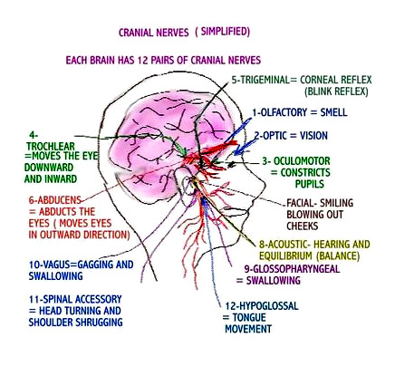
I have both peripheral and cranial (brainstem) neuropathies with myoclonic seizures. I take anti-seizure meds (tegretol) that help control them. Trigeminal neuralgia is considered to be the most painful condition known to man. There is no cure for nerve damage.

The LFA has some of the best information on lupus and neuropathy. From http://www.lfa.org:
The Nervous System

Lupus is an autoimmune disease that can affect almost any part of your body, including your joints, skin, kidneys, heart, lungs, or blood. Lupus can also affect the nervous system and brain. There are several terms doctors use to describe this: neuropsychiatric lupus (NP-SLE), neurocognitive dysfunction, or central nervous system lupus (CNS lupus). Your nervous system has three parts, any of which may be affected by lupus.
The central nervous system (CNS) consists of the brain and the spinal cord.
The peripheral nervous system (PNS) is a network of nerves that connects the brain and spinal cord to the rest of the body, and gives skin and muscles the signals needed for sensation and movement.
The autonomic nervous system (ANS) allows communication between spinal and peripheral nerves and the brain and internal organs, and controls functions like breathing, blood flow, and heart rate.

People with lupus can experience a number of complications when their nervous system is affected. The symptoms may come on suddenly or may come and go, but they will vary depending upon the location and extent of the tissue injury. These symptoms also can be present in other diseases, so diagnosing lupus-related nervous system disorders is often difficult.

Neurologists are the physicians who specialize in the nervous system. They may rely on a number of diagnostic tools to determine whether lupus is involved in your cognitive problems:
x-rays
brain scans (magnetic resonance imaging (MRI) and computed tomography (CT)
electroencephalograms (to capture the electrical pattern of brain activity)
spinal tap (to examine fluid in the spinal column)

Behavioral and cognitive tests may also be done to find out if your memory or other mental abilities have been affected.

Depending on the symptoms, a variety of medications are available to treat lupus-related nervous system disorders, including non-steroidal anti-inflammatory drugs, antimalarials, and steroids. Your response to treatment may be rapid or gradual over several months. For many people with lupus, nervous system involvement is completely reversible.
Central Nervous System (CNS)

When lupus affects your central nervous system, many symptoms may occur, including:
headaches
confusion
fatigue
depression
seizures
strokes
vision problems
mood swings
difficulty concentrating

Drugs used to treat lupus can cause side effects that are similar to the symptoms of CNS lupus. If you have symptoms of CNS lupus you should consult a neurologist who can determine which symptoms are side effects of medication and which are due to lupus. The drugs most known for causing symptoms like those of CNS lupus are:
Non-steroidal anti-inflammatory drugs (NSAIDs) – may cause headache, dizziness, confusion, and in rare instances, meningitis-like symptoms
Antimalarials – in very high doses (not usually given for lupus) may cause manic behavior, seizures, psychosis
Corticosteroids – may cause agitation, confusion, mood swings, psychosis, depression
Anti-hypertensive medications – may cause depression or loss of sex drive

A serious form of lupus called CNS vasculitis may occur when there is inflammation of the blood vessels of the brain. Characterized by high fevers, seizures, psychosis, and meningitis-like stiffness of the neck, CNS vasculitis is the most dangerous form of lupus involving the nervous system and usually requires hospitalization and high doses of corticosteroids to suppress the inflammation.
Peripheral Nervous System (PNS)

The nerves of your peripheral nervous system control your motor responses and sensation, so symptoms of numbness or tingling, or inability to move a part of your body, may be the result of lupus affecting these nerves. Known as peripheral neuropathies, symptoms of PNS nerve damage are caused by inflammation of the nerves or by compression of the nerves due to swelling in the tissue around them. The types of symptoms you might experience include:
vision problems
facial pain
ringing in the ears
dizziness
drooping of an eyelid
carpel tunnel syndrome
Autonomic Nervous System (ANS)

The autonomic nervous system regulates many of your body’s functions that happen almost automatically: heart rate, blood pressure, feeling hot or cold, bladder and bowel functions, release of adrenalin, breathing, sweating, and muscle movement. Lupus can cause these nerve signals to be overactive, which can lead to a wide range of symptoms:
numbness
burning
tingling
mental confusion
headaches
gastrointestinal problems such as nausea, vomiting, constipation, or diarrhea

Raynaud’s phenomenon

Raynaud’s phenomenon is a condition of ANS involvement caused by inflammation of nerves or blood vessels. Blood vessels in your hands and feet go into spasm and restrict blood flow, usually as a reaction to cold temperatures, with the tips of the fingers or toes turning red, white, or blue. Raynaud’s can also cause pain, numbness, or tingling in fingers and/or toes. People who have Raynaud’s phenomenon are advised to avoid cold conditions when possible, and may have to wear gloves or mittens when in air-conditioned surroundings.

Livedo reticularis and palmar erythema are two other skin disorders that may affect you if you have autonomic nerve damage. Both of these conditions can cause a bluish, lacelike mottling under your skin, especially on your legs, giving your skin a “fishnet” look.

Cognitive Dysfunction

As many as half of all people with lupus describe feelings of confusion, fatigue, memory loss, and difficulty expressing their thoughts. This collection of symptoms is termed cognitive dysfunction, although many people call it “lupus fog.”

Cognitive dysfunction most often affects people with mild to moderately active lupus. The causes of these symptoms, and the reasons that the symptoms tend to come and go, are not known. Living with cognitive dysfunction can be very frustrating. However, you can learn to improve your concentration and lessen confusion and memory loss with a variety of coping skills, including puzzles, games, biofeedback, using a daily appointment calendar, and balancing daily activities to reduce stress.

Lupus Headache

Compared with the general population, people with lupus may be twice as likely to experience migraine-like lupus headaches, commonly known as lupus headaches. The features of lupus headaches are similar to migraines and may be seen more often in people who also have Raynaud’s phenomenon. However, headaches can also be caused by vasculitis, a symptom of active lupus due to inflammation of the blood vessels. If you are experiencing headaches that are not improved by an over-the-counter headache medication, be sure to tell your doctor.

Wednesday, April 3, 2013

LUPUS from the Bowels

& Inflammatory Arthritis & Sacroiliitis

From the bowels means just what it sounds like, a little TMI here, but it's part of some our experiences with lupus. Do you have Inflammatory bowel syndrome? Not quite the same as IBS (irritable bowel syndrome) but a PITA just the same! (LOL - PITA, get it?)

Some of the symptoms are constipation, inability to empty your bowels, bloating, gas, nausea and diarrhea. If you do, and you have lupus like I do it can lead to inflammatory arthritis! I did not know this and I am on my second terrible bout of the beast. I've been living on promethazine for nausea for many years.


Perhaps because I also have interstitial cystitis also and a 8mm slipped vertebrae at L4/L5 picking up some inflammatory arthritis in the si joint was a perfect storm.
I'll tell ya (and you can look at my earlier blogs from Dec during my worst ever attack of sacroillitis-it is P A I N FU L!!!!!! I couldn't walk without help, get up or down, sleep on either side, was hot to the touch above my buttocks, my stomach was upset, my pelvis hurt, my legs ached. It's just awful! My grandmother who had undiagnosed lupus and RA had ankylosing spondylitis and it was so severe she was hunchbacked. My poor grandmother, us ungrateful grandkids nicknamed her the Buzzard, how AWFUL! I'm sorry Grandma (looking up at heaven)...

I found some great info on sacroillitis and inflammatory arthritis caused by inflammation from bowel dysfunction-which is common in lupus!  Here it is from: http://www.orthop.washington.edu/?q=patient-care/articles/arthritis/inflammatory-bowel-disease.html

Basics of inflammatory bowel disease

Arthritis means inflammation of joints. Inflammation is a body process that can result in pain swelling warmth redness and stiffness. Sometimes inflammation can also affect the bowel. When it does that process is called inflammatory bowel disease (IBD). IBD is actually two separate diseases: Crohn's disease and ulcerative colitis.
Prognosis

With proper treatment most people who have these diseases can lead full active lives. Usually the inflammation of joints in IBD lasts only a short time and does not cause permanent deformity. With the bowel symptoms under control through medication and diet the outlook for the joints is excellent. 

Incidence
Both men and women are affected equally. The arthritis of IBD can appear at any age but is most common between the ages of 25 and 45.
Joint inflammation begins most often when the colon (the large intestine) is involved in the disease process. In adults the arthritis is usually most active when the bowel disease is active. Indeed the amount of bowel disease usually influences the severity of the arthritis. In children the arthritis is not as often associated with increased bowel disease activity.
Symptoms
Ulcerative colitis
Ulcerative colitis produces inflammation and breakdown along the lining of the colon (see figure 1). Inflammation usually begins in the rectum and extends up the colon. Symptoms may include rectal bleeding abdominal cramping weight loss and fever.
The bowel symptoms often occur before the symptoms of arthritis. When ulcerative colitis is present the arthritis is most likely to occur if there is severe bleeding or if the area around the anus is inflamed. When only the rectum is involved the chance of getting arthritis is less.
Most of the time the arthritis flares (becomes worse) when the bowel symptoms flare. An exception is during the first episode of arthritis which can come at any time. One or more joints may be affected and the symptoms often move from joint to joint. The hips knees and ankles are involved most often although any joint may be affected. The joints may be very painful red and hot but these symptoms usually do not result in permanent damage.
About one-fourth of people with IBD who develop arthritis have a skin rash on the lower legs frequently seen when the arthritis flares. One characteristic rash usually consists of small reddish lumps which are very painful to the touch. This skin condition is called erythema nodosum.
People with ulcerative colitis can develop another form of arthritis called ankylosing spondylitis which involves inflammation of the spine. It usually begins around the sacroiliac joints at the bottom of the back (see figure 2). Symptoms of spondylitis generally do not accompany bowel symptoms in ulcerative colitis. If just the sacroiliac joints are inflamed the symptoms are fairly mild. When the spine is affected however it may be quite painful and even disabling. This can result in stiffness or rigidity.
Crohn's disease
Crohn's disease usually involves either the colon or the ileum the lower small intestine. It may affect both or any part of the digestive tract from the mouth to the rectum. The inflammation involves all layers of the intestinal wall and may lead to scarring and narrowing of the bowel. Fever weight loss and loss of appetite are common symptoms of Crohn's disease.
The arthritis of Crohn's disease can occur before after or at the same time as the bowel symptoms. As with ulcerative colitis the large joints such as the knees and ankles are generally affected though not necessarily on both sides of the body and back pain can result from ankylosing spondylitis.
Causes
The cause of inflammatory bowel disease is not known. Research suggests that the immune system the body's natural defense against foreign invaders is somehow altered in people with these conditions. Researchers believe that the chronic (long-lasting) inflammation present in the intestines of persons with both forms of IBD damages the bowel. This may permit bacteria to enter the damaged bowel wall and circulate through the bloodstream. The body's reaction to this bacteria may then cause problems in other areas of the body. The most common is inflammation of the joints. Other problems include skin sores inflammation of the eyes and certain types of liver disease.
Diagnosis
The history taken by the doctor is the most important part of the diagnosis. Certain information--such as the way the arthritis began the specific joints involved and the relationship between joint and bowel symptoms--is very helpful for diagnosis. The appearance of the joints their range of motion and pain or tenderness during the physical examination are also important. Usually X-rays of the joints are normal unless the joints of the spine are affected. Then damage is visible in X-rays. A blood test for the presence of a substance called HLA-B27 in the blood cells is sometimes helpful in diagnosing ankylosing spondylitis. This substance is an inherited factor present in a much higher frequency among people who have IBD and spondylitis than in the normal population.
Treatment Usually these conditions are treated with medication exercise and sometimes surgery.
Health care team
A gastroenterologist (specialist in diseases of the digestive tract) is usually the doctor who directs treatment but an arthritis or skin specialist may be needed as well.
Diet
Your doctor may give you a special diet to help control your bowel disease. If so follow it carefully. Control of your bowel disease may also help your arthritis. Many diets are advertised as arthritis "cures." There is no known diet that can cure arthritis caused by IBD.
Exercise and therapy
Your doctor or physical therapist will probably design a program of exercises for you to follow every day. Proper exercise helps to reduce stiffness maintain joint motion and strengthen the muscles around the joints. Maintaining the range of motion of affected joints is important in order to prevent or reduce deformity caused by lack of use. If you have ankylosing spondylitis range of motion exercises of the spine are of benefit. Deep breathing exercises are emphasized because motion of the ribs may eventually be restricted as the disease moves up the spine. If you smoke you should stop in order to help prevent breathing complications.
If you find exercising to be painful take a warm shower or bath before you exercise. This should lessen the pain and stiffness. Begin the exercises slowly and plan them for the times of the day when you have the least pain.
Good posture is essential for the person with ankylosing spondylitis and IBD. The spine should be kept as straight as possible at all times. Avoid sitting for prolonged periods of time. Sleep on your stomach or back on a firm mattress. If you need to use a pillow under your head only use a thin one or one that fits the hollow of your neck. Avoid pillows under your knees. Keep your body as straight as you can. Avoid lying in a curled position.
Medications
Several medications may be helpful in controlling arthritis and IBD. Sulfasalazine is a very useful sulfa drug. The other medications fall into certain groups of drugs: corticosteroids, immunosuppressives and nonsteroidal anti-inflammatory drugs (NSAIDs).
Sulfasalazine (Azulfidine) helps to control both the bowel disease and the symptoms of arthritis. It is usually started at a low dose to lessen possible side effects and then increased if needed. The most common side effects are nausea and headaches. The nausea may be controlled by taking the drug with food or by using the enteric-coated form of the drug. (This form is specially designed to dissolve in the bowel not in the stomach.)
Sulfasalazine can usually be taken safely for a long time. Some people however develop an allergy to sulfasalazine in the form of a rash and fever. Giving the drug in frequent very small doses may enable the person to tolerate the drug without producing a rash or other reaction. When sulfasalazine cannot be taken due to side effects or allergy olsalazine (Dipentum) or mesalamine (Asacol) may be taken but these drugs have not been shown to be effective against arthritis.
Corticosteroids are similar to cortisone a hormone produced by the body. They are strong anti-inflammatory drugs which can help both the symptoms of the bowel and the joints. They are used only when the symptoms are severe because they may produce serious side effects when taken for a long time. These side effects include thinning of bones (osteoporosis) cataracts reduced resistance to infection diabetes obesity and high blood pressure.
Be sure to discuss the possible side effects with your doctor before taking corticosteroids. Most of the side effects decrease and eventually go away as the dosage is reduced and stopped. Once you begin taking these drugs however never stop or change the dosage on your own.
Immunosuppressives such as azathioprine (Imuran) are used on occasion for arthritis and Crohn's disease. By suppressing the immune system they reduce inflammation. The most common side effect of these medications is a decrease in white blood cells which can cause an increased risk of infections. Other side effects of these medications may include fever rash vomiting hair loss and liver toxicity. Immunosuppressives therefore are used with caution.
Nonsteroidal anti-inflammatory drugs (NSAIDs) such as ibuprofen are helpful in controlling the pain swelling and stiffness of inflamed joints. To work effectively they must be taken every day during the arthritis episode.
NSAIDs may produce nausea indigestion and heart burn. In addition they may cause bleeding from the stomach and make the underlying bowel disease worse so they are used with caution in IBD. These side effects can usually be decreased if the drug is taken with food fluid or an antacid.
Surgery
Surgical removal of the diseased bowel is usually a permanent cure for ulcerative colitis. This surgery also puts an end to any arthritis that may be present unless the arthritis involves the spine. Ankylosing spondylitis may last even after removal of the diseased colon.
Crohn's disease does not respond as well to surgery. Surgical removal of the diseased bowel may be necessary but it does not cure Crohn's disease. Thus symptoms of arthritis may recur when and if bowel symptoms reappear.
Strategies for coping
Living with arthritis and IBD can be very difficult at times. In addition to pain and discomfort you may have to deal with changes in your appearance or in your leisure time activities. These changes may leave you sad depressed or angry. Relaxation techniques are coping skills that can help you relieve pain and stress and adjust to the changes in your life.
It helps to talk about your feelings with family, members friends or someone else who has arthritis and IBD. Ask your doctor about educational programs materials or support groups for people who have arthritis as well as their families.
Resources
Another source of help is the Crohn's and Colitis Foundation of America Inc. (CCFA). It provides educational materials and programs for people who have IBD. To locate the chapter nearest you contact the CCFA at info@ccfa.org write to them at 386 Park Avenue South 17th Floor New York NY 10016-8804 or call toll-free (800) 932-2423.
Credits
Some of this material may also be available in an Arthritis Foundation brochure. Contact the Washington/Alaska Chapter Helpline: (800) 542-0295. If dialing from outside of WA and AK contact the National Helpline: (800) 283-7800.