Tuesday, September 9, 2014

Learning how to not be "RASH"! My pemphigus/pemphigoid story-

How skin falls apart: The pathology of autoimmune skin disease is revealed at the nanoscale - UoB study of pemphigoid rashes discovered new details of how autoantibodies destroy healthy cells in skin! 

This information provides new insights into autoimmune mechanisms in general and could help develop and screen treatments for patients suffering from all autoimmune diseases, estimated to affect 5-10 percent of the U.S. population.

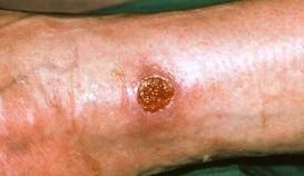
The study of pemphigoid rashes is of interest to people like me, who get these types of rashes-here's a pic of me with one-
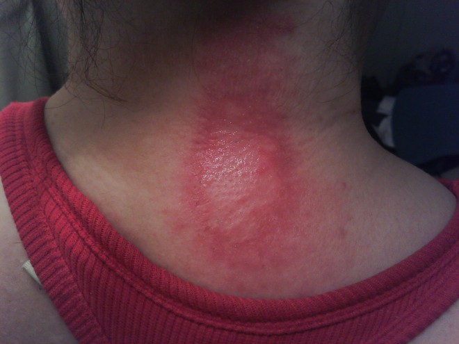
My Pemphigoid Rash
My Pemphigoid Rash
It hurt, felt like my skin was pulling, then it seeped clear white blood cells.  I could actually smell them!  (Yuk, right?)  It took quite a while to heal completely.  It took about 5 months to completely heal.  It did not leave any discolored skin. I used betamethazone to heal it.  A stronger type of steroid cream &/or a steroid shot right into the rash would have probably healed it faster, but I didn't know that then.  I had this rash Feb 2013. It popped in right after a radiofrequency ablation to my cervical spine for cervical spondylitis & radiculopathy right at the shot site!  Imagine that-Lupus stopping by to say hello just because I had a shot there-(how nice....NOT!  lol)
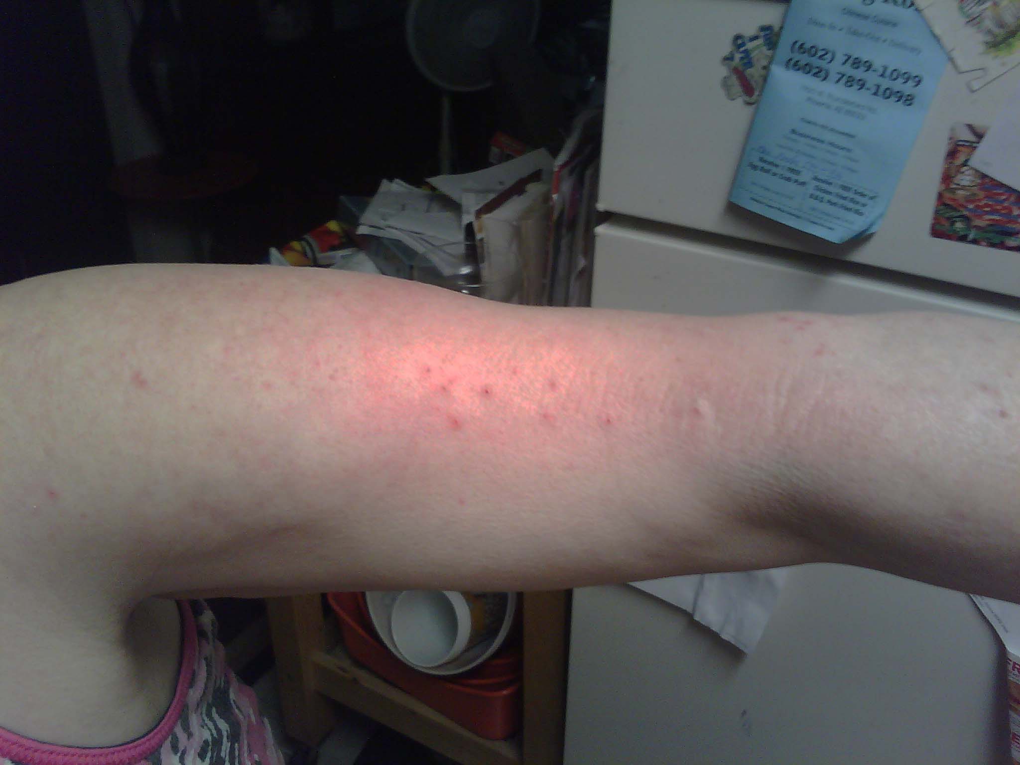
I've only had one pemphigoid type rash since, and it was a small one.  Since being treated for lupus with Cellcept (mycophenalate) my rashes (discoid, pemphigoid, porphyria, urticaria) have all been minimal.  Much less severe!!!  Of course now I stay in as much as possible, it's the only real way to minimize the rashes.  Don't go out during the day!
Here's some info on Pemphigoid Rashes!

Definition

Pemphigoid is a group of subepidermal, blistering autoimmune diseases that primarily affect the skin, especially the lower abdomen, groin, and flexor surfaces of the extremities. Here, autoantibodies (anti-BPA-2 and anti-BPA-1) are directed against the basal layer of the epidermis and mucosa.
The condition tends to persist for months or years with periods of exacerbation and remission. Localized variants of the condition have been reported, most often limited to the lower extremities and usually affecting women.

Types

There are two predominant types of pemphigoid: mucous membrane pemphigoid (MMP) also called cicatricial pemphigoid, and bullous pemphigoid (BP) (g). Pathogenesis and management are quite different for these conditions. Scar formation in mucous membrane pemphigoid can lead to major disability (g).

Pemphigoid gestationis

Pemphigoid gestationis (PG) is a rare autoimmune bullous dermatosis of pregnancy. The disease was originally named herpes gestationis on the basis of the morphological herpetiform feature of the blisters, but this term is a misnomer because PG is not related to or associated with any active or prior herpes virus infection (h). PG typically manifests during late pregnancy, with an abrupt onset of extremely pruritic urticarial papules and blisters on the abdomen and trunk, but lesions may appear any time during pregnancy, and dramatic flares can occur at or immediately after delivery. PG usually resolves spontaneously within weeks to months after delivery.

Epidemiology

Bullous Pemphigoid:

  • BP is the most frequent blistering disease of the skin (and mucosa) affecting typically the elderly (>65 years) (k), but can occur at any age and in any race.
  • Overall incidence: ± 7-10 new cases per million inhabitants per year.
  • After the age of 70 incidence significantly increases.
  • Relative risk for patients > 90 y have a 300-fold higher than those < 60 y.
  • Women and men equally afflicted.
  • Precipitating factors include trauma, burns, ionizing radiation, ultraviolet light, and certain drugs such as neuroleptics and diuretics, particularly lasix (furosemide), thiazides, and aldosterone antagonists.
  • Correlations between BP flare activity and recurrence of underlying cancer suggest such an association in some patients.
  • Even without therapy, BP can be self-limited, resolving after a period of many months to years, but is still a serious condition especially in the elderly.
  • (j) 1-y survival probability may be as high as 88.96% (standard error 5.21%), with a 95% confidence interval (75.6%, 94.2%) but other analyses have documented 1 year mortalities of as much as 25-30% in moderate to severe pemphigus even on therapy (hh).
  • Genetics
    • Genetic predisposition, but not hereditary

Pemphigoid Gestationis:

  • Is a condition of pregnancy (childbearing age women).
  • In the United States, PG has an estimated prevalence of 1 case in 50,000-60,000 pregnancies.
  • No increase in fetal or maternal mortality has been demonstrated, although a greater prevalence of premature and small-for-gestational-age (SGA) babies is associated with PG.
  • Patients with PG have a higher relative prevalence of other autoimmune diseases, including Hashimoto thyroiditis, Graves disease, and pernicious anemia.

Histology

The earliest lesion of BP is a blister arising in the lamina lucida, between the basal membrane of keratinocytes and the lamina densa. This is associated with loss of anchoring filaments and hemidesmosomes. Histologically, there is a superficial inflammatory cell infiltrate and a subepidermal blister without necrotic keratinocytes. The infiltrate consists of lymphocytes and histiocytes and is particularly rich in eosinophils. There is no scarring.
Approximately 70% to 80% of patients with active BP have circulating antibodies to one or more basement membrane zone antigens.
  • Autoantibodies to BP180 (and BP230).
  • BP180 and BP230 are two components of hemidesmosomes, junctional adhesion complexes.
  • T cell autoreactive response to BP180 and BP230 regulate autoantibody production (l).
  • On direct immunofluorescence, the antibodies are deposited in a thin linear pattern; and on immune electron microscopy, they are present in the lamina lucida. (By contrast, the antibodies to basement membrane zone antigens that are present in cutaneous lupus erythematosus are deposited in a granular pattern).

Clinical Features

Pemphigoid

Bullous Pemphigoid (BP) is subepidermal blistering autoimmune disease primarily affects the skin, especially the lower abdomen, groin, and flexor surfaces of the extremities. Mucous membrane involvement is seen in 10%-40% of patients. The disease tends to persist for months or years with periods of exacerbation and remission.
The spectrum of presentations is extremely broad (l), but typically there is an itchy eruption with widespread blistering, and tense vesicles and bulla (blisters), with clear fluid (can be hemorrhagic) on apparently normal or slightly erythematous skin.
Lesions normally appear on the torso and flexures, particularly on the inner thighs. Blisters can range in size from a few millimeters to several centimeters, and although. pruritic, typically heal without scarring.
Sometimes erosions and crusting is seen. Also itchy bumps (papules) and crusts (plaques) can be seen with an annular or figurate pattern. A characteristic feature is that multiple bullae usually arise from large (palm-sized or larger), irregular, urticarial plaques. (r) Mucosal (oral, ocular, genital) involvement is also sometimes present, but ocular involvement, is rare. (s) BP can be difficult to diagnose in its ‘non-blistering’ stage, when just itchy, red, elevated patches are visible. Erosions are much less common than in pemphigus, and the Nikolsky sign is negative.
BP is characterized by spontaneous remissions followed by flares in disease activity that can persist for years. Even without therapy, BP is often self-limited, resolving after a period of many months to years, but may become very extensive.
Localized variants of the disease have been reported, most often limited to the lower extremities and usually affecting women. One such variant, localized vulvar pemphigoid, reported in girls aged 6 months to 8 years, presents with recurrent vulvar vesicles and ulcerations that do not result in scarring (t)
Bullous pemphigoid is distinguished from other blistering skin diseases, such as linear IgA dermatosis, epidermolysis bullosa acquisita, and cicatricial pemphigoid, by the following 4 clinical items (it can also be distinguished by biopsy and certain immunological tests) (u)
  • Absence of atrophic scars;
  • Absence of head and neck involvement;
  • Relative absence of mucosal involvement.

Mucous membrane pemphigoid

Mucous membrane pemphigoid (MMP) is a chronic autoimmune disorder characterized by blistering lesions that primarily affect the various mucous membranes of the body, but also affects the skin (MMP is now the preferred term for lesions only involving the mucosa) (v). It is also known as Cicatricial Pemphigoid (CP), as it is often scarring.
The mucous membranes of the mouth and eyes are most often affected, but those of the nose, throat, genitalia, and anus may also be affected. The symptoms of MMP vary among affected individuals depending upon the specific site(s) involved and the progression of the disease. Disease onset is usually between 40 and 70 years and oral lesions are seen as the initial manifestation of the disease in about two thirds of the cases. Blistering lesions eventually heal, sometimes with scarring. Progressive scarring may potentially lead to serious complications affecting the eyes and throat.
There is no racial or ethnic predilection although most studies have demonstrated a female to male ratio of approximately 2:1 (w). The diagnosis of MMP is mainly based on history, clinical examination and biopsy of the lesions.
More on Pemphigoid Rashes from the Pemphigus & Pemphigoid Foundation HERE!

MORE HERE from the University of Buffalo-

Friday, August 29, 2014

Leave Your Ego At the Door & Join/Share at "The Lupus Channel"!

lupuschannelcommunity
Information is Power!  
My goal at "The Lupus Channel" Google community is to share all things Autoimmune-
Articles, Info, Blogs, Events, Videos & Vlogs, Webinars, Pinterest Boards, Support Groups, Research & Studies, etc on: Lupus Fibromyalgia Sjogrens Scleroderma MCTD (mixed connective tissue disease  & other autoimmune conditions
AND to help you connect to other people just like YOU & Me who learn from each other everyday by sharing experiences on how to most effectively manage our disease and have quality of life!  Nice to meet you & WELCOME!  JJ
A Special Note to Advocates:  Feel FREE to share your links to your groups, your posts & your contact on "The Lupus Channel"-I left my ego at the door- the objective is simply to reach the patients who need help!  TY so much!  I look forward to your posts!
Click HERE to JOIN "The Lupus Channel: Google Community-

My goal is to share all things Autoimmune-Articles, Info, Blogs, Events, Videos & Vlogs, Webinars, Pinterest Boards, Support Groups, Research & Studies, etc on:
  • Lupus
  • Fibromyalgia
  • Sjogrens
  • Scleroderma
  • MCTD (mixed connective tissue disease
  •  & other autoimmune conditions
And to help you connect to other people just like YOU & Me who learn from each other everyday by sharing experiences on how to most effectively manage our disease and have quality of life!
Nice to meet you!  JJ
ps. I make lupus music videos to help spread awareness so that we can get closer everyday to a cure!  Here are my videos- HERE!
Hook up with The Lupus Channel on Twitter HERE!  And subscribe to my Blog HERE!
Also Please Join The Lupus Channel FB Group HERE!
My Personal Accounts
You can "Friend" me on FB- HERE!  And you can "Follow" me on Twitter- HERE!
Also-feel free to email me anytime HERE!
Your not alone with lupus or any autoimmune disease-there is tons of help out here!  Reach out TODAY!

Wednesday, August 20, 2014

Rheums with a View: Rheumatologists Point of View at Patients Requesting Tests

meddiseasescartoon
Article from "The Rheumatologist" official publication of the ACR
by Matthew J. Koster, MD, & Kenneth J. Warrington, MD
Case
A 28-year-old woman presents for evaluation of exhaustion, widespread myalgias and muscle spasms. In addition, she has numerous symptoms spanning multiple organ systems, including paresthesia, atypical chest pain and abdominal bloating. She has previously undergone evaluation at other medical centers and by multiple subspecialists, and no specific pathology or diagnosis has been established. She has difficulty continuing her employment due to disabling symptoms. A compre­hensive evaluation for malignancy and infectious diseases has been unrevealing. Neurological evaluation, including detailed physical examinations, imaging of the brain, electromyography and testing for large- and small-fiber neuropathy, has been negative. Extensive laboratory studies, autoimmune serologies and radiographic imaging studies are unremarkable.
You review the history and conduct a comprehensive physical examination and find no abnormalities, except for widespread muscle tenderness without weakness. The patient is not reassured by the negative evaluation and is concerned that a progressive auto­immune disorder is being missed. Additional evaluation is pursued, including magnetic resonance imaging (MRI) of proximal muscles, which is negative. The patient requests further testing, including a muscle biopsy. Her request conflicts with your recommendation that the diagnostic evaluation has been completed and your plan to pursue symptomatic management for fibromyalgia.
Dilemma
As access to medical information expands, patients increasingly present to clinical encounters with specific preconceptions regarding their possible diagnosis and requests for testing and interventions. Medical literacy, if applied appropriately, can lead to patient education and empowerment, and can improve shared decision making. However, if the information obtained is of questionable accuracy, incomplete, misinterpreted or outdated, such requests and expectations can lead to challenging clinical encounters, especially when the patient’s preconceptions differ from the physician’s assessment.
How should physicians approach patient requests for testing deemed unnecessary or even contraindicated in a manner that respects patient preferences, upholds a physician’s integrity and maintains a strong physician–patient relationship?
Discussion
On the surface, the patient’s request in the above scenario may seem morally innocuous; however, it has complex ethical undertones involving patient autonomy, physician professionalism, knowledge asymmetry, medical uncertainty and defensive medicine.
Should additional testing be performed in this patient?
The patient in this clinical scenario has fibromyalgia. Comprehensive examination and testing have already been performed to rule out other conditions for the cause of pain, one of the criteria for the diagnosis of this condition.1 Muscle biopsy studies show there are no specific changes conclusive for fibromyalgia.2 Additional testing in the evaluation of such patients should be prompted primarily by clinical sus­picion and objective physical findings. Excessive and repeated testing may have a negative effect on the patient’s well-being and encourage medicalization.3 In the absence of findings concerning for inflammatory myopathy, muscular dystrophy or metabolic muscle disease, muscle biopsy would not be indicated and, thus, can be considered medically unnecessary in this case.
Matthew Koster
Matthew Koster
What are positive & negative rights?
When patient requests conflict with a physician’s concept of acceptable practice, it’s important to examine the distinction between positive and negative rights.4 When considering medical encounters, most ethicists focus on patient autonomy in terms of negative rights—that is, the right to decline a treatment or test.
The American Medical Association Code of Ethics states the patient has the right to make decisions regarding the healthcare that is recommended by his or her physician. Accordingly, patients may accept or refuse any recommended medical treatment.5 The emphasis of informed consent promotes appropriate barriers to prevent patients from receiving care or interventions that are contrary to their desires or beliefs. Respect for patient autonomy, however, also governs positive rights—the right that something be done. Much less guidance is provided to physicians in this regard. If physicians are to respect patient autonomy by way of positive rights, how are physicians ever justified in denying a patient’s request?
According to the ACP Ethics Manual, the patient–physician relationship entails special obligations for the physician to serve the patient’s interest because of the specialized knowledge that physicians possess, the confidential nature of the relationship, and the imbalance of power between patient and physician.6
In this case scenario, the patient’s autonomous decision conflicts with the physician’s professional duty to look out for the patient’s best interests and welfare (i.e., beneficence). Therefore, we must carefully consider the balance between respecting the patient’s right to share in the medical decision making and the physician’s responsibility to avoid patient harm (i.e., non­maleficence). This patient is so worried about having an autoimmune disease that her quality of life is com­promised. When patients request diagnostic studies that are not indicated, physicians should first seek to understand the reason for the request. Then, the physician should educate the patient about his or her rationale for not recommending the test based on the physician’s specific knowledge and medical expertise for which the patient is seeking counsel.
When the risk of harm to the patient significantly exceeds the potential diagnostic utility of an intervention, the physician has to keep in focus one of the principal precepts of bioethics: primum non nocere (first, do no harm).
Kenneth Warrington
Kenneth Warrington
What if, despite education of medical necessity & risk-benefit ratio of intervention, the patient continues to insist on the nonindicated intervention?
If the principles of patient autonomy, beneficence and nonmaleficence were the only factors being weighed in such encounters, physicians may convince themselves that acquiescence to patient requests can be justified on grounds that reduction of anxiety alone may tip the risk-benefit scale toward intervention.
However, in the current case, it seems doubtful that another negative test would be the end of her requests. In addition, it is increasingly apparent in our current healthcare environment that medically unnecessary testing cannot be sustained, and the ethical tenet of distributive justice must also be considered. The principle of distributive justice implies that healthcare resources should be distributed fairly in society. Therefore, although an additional laboratory test, imaging study or procedure may seem insignificant, when magnified on a national scale, the economic cost of these “inconsequential” decisions becomes large. It is not unethical to withhold nonindicated treatment, and one of the reasons is the pursuit of distributive justice.
We must carefully consider the balance between respecting the patient’s right to share in the medical decision making & the physician’s responsibility to avoid patient harm.
But what if I decline a test & miss something diagnosable?
Societal, legal and professional pressures add to physicians’ fear of missing a diagnosis. This is compounded in disorders when symptoms are purely subjective. When dealing with medical uncertainty associated with vague or subjective symptoms, physicians may agree to patient requests that do not present undue risk to the patient. However, the testing should be rational, medically justified and part of a comprehensive, well-documented management plan.
Back to the Patient
A critical goal of this patient’s physician is to reassure her that there is not an underlying disease being missed. The assumption in this encounter is that the patient feels information that can be obtained from a biopsy is more convincing than the physician’s clinical judgment regarding her diagnosis.
Additional investigation into the patient’s request reveals some important information: A friend was recently diagnosed with polymyositis, and she is concerned she may have this as well. After review of her current workup, education about fibromyalgia and its differences from autoimmune disorders, as well as the risk of the procedure compared with the expected benefit, the patient ultimately agrees a muscle biopsy is not required. Although she is still apprehensive about her diagnosis, she agrees to initiate a treatment plan with the reassurance that she will have ongoing monitoring for any changes in symptoms and reevaluation as necessary—plans that can only be implemented in the context of a strong physician–patient relationship.
Have you had an experience in which patients requested tests or treatment that you felt was not indicated? How did you handle the request? If you have comments or questions about this case, or if you have a case that you’d like to see in Ethics Forum, e-mail us at klosavio@wiley.com.
Matthew J. Koster, MD, is a second-year rheumatology fellow at Mayo Clinic, Rochester, Minn.
Kenneth J. Warrington, MD, is an associate professor of medicine and practice chair of rheumatology at Mayo Clinic, Rochester, Minn. He is a member of the ACR’s Committee on Ethics and Conflict of Interest.
Acknowledgment
The authors thank Dr. Robert H. Shmerling, MD, for his critical review of the manuscript.
References
Wolfe F, Clauw DJ, Fitzcharles M-A, et al. The American College of Rheumatology preliminary diagnostic criteria for fibromyalgia and measurement of symptom severity. Arthritis Care Res. 2010 May;62(5):600–610.
Bengtsson A. The muscle in fibromyalgia. Rheumatology. 2002 Jul;41(7):721–724.
Fitzcharles M-A, Ste-Marie PA, Goldenberg DL, et al. Canadian Pain Society and Canadian Rheumatology Association recommendations for rational care of persons with fibromyalgia. A summary report. J Rheum. 2013 Aug;40(8):1388–1393.
Beauchamp TL, Childress JF. Principles in Bioethics. New York: Oxford University Press. 1979:50.
AMA Council on Ethical and Judicial Affairs. Code of Medical Ethics of the American Medical Association, 2012–2013.
Snyder L. American College of Physicians Ethics, Professionalism, and Human Rights Committee. American College of Physicians Ethics Manual, 6th Ed. Ann Intern Med. 2012 Jan 3;156(1 Pt 2):73–104.
http://www.the-rheumatologist.org/details/article/6480961/Patient_Test_Requests_Pose_Challenges_for_Rheumatologists.html

Monday, August 4, 2014

The Inflammation connected to the Hamstring..The Hamstring connected to the Tendon..& that’s the way of the Lupus!

myleg
hamstring tendonitis from lupus-inflammation
So-there it is.  Tendonitis in the hamstring.  NO, I did not fall even though YES, I am a klutz.  And NO, I did not get punched! (except by lupus)

I woke up a couple days ago and Surprise- OWWWWWWWWWWWWWWWWWWWWWWWWW!  It's hot, swollen and yes, painful.  It's a pulling type of pain, from around my calfs up to my lower thighs and back of my leg.  My doc is scheduling a cortisone shot which will deflate the inflammation and the pain asap.  I'm also taking steroids and anti-inflammatories.  It helps.  Alot.  Ice is indicated for inflammation as well.
I tried to get a selfie of this but ended up tangled and unbalanced like you get playing Twister!  Finally got hubbie to snap a photo-it even shocks ME & it's on my body!
So for those who question what kind of inflammation mixed connective tissue disease causes- tendonitis is just one of em!  Lupus is a mixed connective tissue disease-and our bodies are made of this material-so it's all up for lupus damage grabs, lol.  Two days of bedrest and a few shots isn't so bad-

"Housebound is no worse than earthbound it's what you make of it."

Here's some more info on hamstring tendonitis & lupus connective tissue damage from Medicine Net:

Connective tissue diseases are actually a group of medical diseases. A connective tissue disease is any disease that has the connective tissues of the body as a primary target of pathology. The connective tissues are the structural portions of our body that essentially hold the cells of the body together. These tissues form a framework, or matrix, for the body. The connective tissues are composed of two major structural protein molecules, collagen and elastin. There are many different types of collagen protein that vary in amount in each of the body's tissues. Elastin has the capability of stretching and returning to its original length -- like a spring or rubber band. Elastin is the major component of ligaments (tissues that attach bone to bone) and skin. In patients with connective tissue diseases, it is common for collagen and elastin to become injured by inflammation.
Many connective tissue diseases feature abnormal immune system activity with inflammation in tissues as a result of an immune system that is directed against one's own body tissues (autoimmunity).

Diseases in which inflammation or weakness of collagen tends to occur are also referred to as collagen diseases. Collagen vascular disease is a somewhat antiquated term used to describe diseases of the connective tissues that typically include diseases that can be (but are not necessarily) associated with blood vessel abnormalities.


Monday, June 2, 2014

SUN is out, Wind in My Face; Absolutely Awful! ~Life Above Zero~


lupusgrumpycat
Not only vampires go out at night-LUPUS patients do too!
While these things are true-UV Hypersensitivity is prevalent with Systemic Lupus Erythematosus - it doesn't have to mean ISOLATION in this day & age!
With lupus, our bodies aren't just "allergic" to itself (white blood cells attacking healthy tissue by mistake causing central nervous system damage, kidney damage, skin damage including hives, solar urticaria, discoid, pemphigoid, and cutaneous porphyria rashes, lesions & vasculitis, inflammation around the heart-pericarditis, around the lungs-pleurisy, blood disorders (clotting issues and anemia), nerve damage and a myriad of other symptoms and conditions.  So what do you do?  You manage.  You CONFORM!

I can tell you what I do-
My windows have double black-out drapes.  I go out mostly at night only.  After many years of neutragena helioplex 360 and 120 proof sunscreen-rashes and lupus flares anyway-I discovered the true way to be less sick is to just plain STAY INSIDE.  In the dark.  No UV lights, no sun at all.  Is it isolating?  I don't think so thanks to modern technology-but that's me-it's all in how you see it.  "Housebound is no worse than earthbound it's what you make of it."
Here's some of my rashes from UV-if you had these I'm sure you'd come to the same conclusion!
discoid-rash-400x400 malarrash - Copy IMG_20130130_164207(4) headrash1 juliefacerash vasculitislegs vascularlesion june2013rashjulieshingles1
That would drive ya inside-yes?  LOL..And my point?  It doesn't have to BE isolating or feel isolating..
The internet has given us many advances-immediate chat, visuals like skype, and the ability to find info, give info, and reach out without having to take one step outside into that debilitating sun.  I can watch my son's chorus concert from bed, talk to people on the other side of the world-make appointments, talk to doctors and do relatively anything but reach out and touch someone!  So isolating?  No-it's the opposite for people like me!!!  It's FREEING!  And I'm thankful for it everyday!  Even after a small TIA June 2012-I was relatively a "head in a bed" while I recouped...but I was happy and I was anything but bored or isolated!  So again-Housebound is what you make of it!  Is a wheelchair a good or a bad thing?  Ask someone who is in one?  It's a great thing!  It's mobility!  Just a different way-and so what!
On a personal note - I enjoy watching reality shows-specifically shows about Alaska-the last frontier.  Why- I wonder?  Because we LUPUS patients share something with these people-we are isolated.  We are literally in the dark most of the time.  Plus of course-they are exciting!  There's gold miners, hunters, and people who are solely responsible for their own food and water sources-and live in the most extreme of circumstances.  And they succeed.  They flourish.  They give us HOPE.  They tell us that life is precious-and to be lived-  There is a woman you might have all seen-Susan Aikens on a National Geographic show called "Life Below Zero", who owns and manages a remote fueling station near the Arctic Circle-she is alone 8 months of the year.  She is the strongest, toughest woman I have ever seen.  She was attacked by a bear, left for dead, sewed up her own head, hunts for food and braves temperatures so freezing- she doesn't just persevere-she flourishes.   I designed the pic of Sue & Alaska to say Thank You for the Inspiration!
lighthousesusanaikens
Pic of Susan Aikens from "Life Below Zero" on National Geographic channel
Susan said that she knows due to her remoteness and the difficulty especially in winter for planes and helicopters to reach her that she and people like her understand that if something happens, if they get hurt-that there is always the chance that noone will come and they will die out there.  People with chronic illness know the same fear.  It is freeing to accept what might happen-what could happen-it takes the fear out of it!  Still-I think Susan has another message-one she might not even realize-
When Susan Aikens was asked why she is alright to stay out there in remote Alaska alone and be isolated so much of the year-she said it is because when she was little all she wanted to be was a lighthouse keeper.  Well-she IS a lighthouse keeper.  She's a beacon of what life without fear means for those with chronic illness.  She's an inspiration.
"We are told to let our light shine, and if it does, we won't need to tell anybody it does. Lighthouses don't fire cannons to call attention to their shining- they just shine"., Dwight L Moody


from Wiki on Solar Urticaria:
Solar urticaria (SU) is a rare condition in which exposure to ultraviolet or UV radiation, or sometimes even visible light, induces a case of urticaria or hives that can appear in both covered and uncovered areas of the skin.[1][2] It is classified as a type of physical urticaria.[3] The classification of disease types is somewhat controversial. One classification system distinguished various types of SU based on the wavelength of the radiation that causes the breakout; another classification system is based on the type of allergen that initiates a breakout.[4][5]
The agent in the human body responsible for the reaction to radiation, known as the photoallergen, has not yet been identified.[6] The disease itself can be difficult to diagnose properly because it is so similar to other dermatological disorders, such as polymorphic light eruption or PMLE.[7] The most helpful test is a diagnostic phototest, a specialized test which confirms the presence of an abnormal sunburn reaction. Once recognized, treatment of the disease commonly involves the administration of antihistamines, and desensitization treatments such as phototherapy.[1] In more extreme cases, the use of immunosuppressive drugs and even plasmapheresis may be considered.[8]
The initial discovery of the disease is credited to P. Merklen in 1904, but it did not have a name until the suggestion of "solar urticaria" was given by Duke in 1923.[6][9] However, their research contributed to the study of this uncommon disease. More than one hundred cases have been reported in the past century.[10]
from the Lupus Foundation of America on Lupus Rashes:
Approximately two-thirds of people with lupus will develop some type of skin disease, called cutaneous lupus erythematosus. Skin disease in lupus can cause rashes or sores (lesions), most of which will appear on sun-exposed areas such as the face, ears, neck, arms, and legs.
40-70 percent of people with lupus will find that their disease is made worse by exposure to ultraviolet (UV) rays from sunlight or artificial light.
A dermatologist, a physician who specializes in caring for the skin, should treat lupus skin rashes and lesions. He or she will usually examine tissue under a microscope to determine whether a lesion or rash is due to cutaneous lupus: taking the tissue sample is called a biopsy.

The Forms of Cutaneous Lupus

Lupus skin disease can occur in one of three forms:
  1. Chronic cutaneous (discoid) lupus
  2. Subacute cutaneous lupus
  3. Acute cutaneous lupus.
Chronic cutaneous lupus (discoid lupus) appears as disk-shaped, round lesions. The sores usually appear on the scalp and face but sometimes they will occur on other parts of the body as well.
Approximately 10 percent of people with discoid lupus later develop lupus in other organ systems, but these people probably already had systemic lupus with the skin rash as the first symptom.
Discoid lupus lesions are often red, scaly, and thick. Usually they do not hurt or itch. Over time, these lesions can produce scarring and skin discoloration (darkly colored and/or lightly colored areas). Discoid lesions that occur on the scalp may cause the hair to fall out. If the lesions form scars when they heal, the hair loss may be permanent.
Cancer can develop in discoid lesions that have existed for a long time. It’s important to speak with your doctor about any changes in the appearance of these lesions.
Discoid lupus lesions can be very photosensitive so preventive measures are important:
  • Avoid being out in the sunlight between the hours of 10 a.m. and 4 p.m.
  • Use plenty of sunscreen when you are outdoors
  • Wear sun-protective clothing and broad-brimmed hats
  • Limit the amount of time spent under indoor fluorescent lights
Subacute cutaneous lesions may appear as areas of red scaly skin with distinct edges or as red, ring-shaped lesions. The lesions occur most commonly on the sun-exposed areas of the arms, shoulders, neck, and body. The lesions usually do not itch or scar, but they can become discolored. Subacute cutaneous lesions are also photosensitive so preventive measures should be taken when spending time outdoors or under fluorescent lights.
Acute cutaneous lupus lesions occur when your systemic lupus is active. The most typical form of acute cutaneous lupus is a malar rash–flattened areas of red skin on the face that resemble a sunburn. When the rash appears on both cheeks and across the bridge of the nose in the shape of a butterfly, it is known as the "butterfly rash." However, the rash can also appear on arms, legs, and body. These lesions tend to be very photosensitive. They typically do not produce scarring, although changes in skin color may occur.

Other Skin Problems

There are several other conditions that can occur with lupus:
Calcinosis is caused by a buildup of calcium deposits under the skin. These deposits can be painful, and may leak a white liquid. Calcinosis can develop from a reaction to steroid injections or as a result of kidney failure.
Cutaneous vasculitis lesions occur when inflammation damages the blood vessels in the skin. The lesions typically appear as small, red-purple spots and bumps on the lower legs; occasionally, larger knots (nodules) and ulcers can develop. Vasculitis lesions can also appear in the form of raised sores or as small red or purple lines or spots in the fingernail folds or on the tips of the fingers. In some cases, cutaneous vasculitis can result in significant damage to skin tissue. Areas of dead skin can appear as sores or small black spots at the ends of the fingers or around the fingernails and toes, causing gangrene (death of soft tissues due to loss of blood supply).
Hair loss can occur for other reasons besides scarring on the scalp. Severe systemic lupus may cause a temporary pattern of hair loss that is then replaced by new hair growth. A severe lupus flare can result in fragile hair that breaks easily. Such broken hairs at the edge of the scalp give a characteristic ragged appearance termed "lupus hair."
Raynaud’s phenomenon is a condition in which the blood vessels in the hands and/or feet go into spasm, causing restricted blood flow. Lupus-related Raynaud’s usually results from inflammation of nerves or blood vessels and most often happens in cold temperatures, causing the tips of the fingers or toes to turn red, white, or blue. Pain, numbness, or tingling may also occur. People with Raynaud’s phenomenon should try to avoid cold conditions, and, if necessary, should wear gloves or mittens and thick socks when in an air-conditioned area.
Livedo reticularis and palmar erythema are caused by abnormal rates of blood flow through the capillaries and small arteries. A bluish, lacelike mottling will appear beneath the skin, especially on the legs, giving a "fishnet" appearance. Like Raynaud’s phenomenon, these conditions tend to be worse in cold weather.
Mucosal ulcerations are sores in the mouth or nose or, less often, in lining of vaginal tissue. These ulcers can be caused by both cutaneous lupus and systemic lupus. It is important to differentiate lupus ulcers from herpes lesions or cold sores, which may be brought on by the use of immunosuppressive drugs. Lupus ulcers are usually painless and signs of inflammation will show up in the biopsy.
Petechiae (pah-TEE-kee-eye) are tiny red spots on the skin, especially on the lower legs, that result from low numbers of platelet in the blood, a condition called thrombocytopenia. Although thrombocytopenia is common in lupus, serious bleeding as a result of the low number of platelets usually does not occur.

Treating Cutaneous Lupus

The medications used to treat lupus-related skin conditions depends on the form of cutaneous lupus. The most common treatments are topical ointments, such as steroid cream or gel. In some cases liquid steroids will be injected directly into the lesions.
A new class of drugs, called topical immunomodulators, can treat serious skin conditions without the side effects found in corticosteroids: tacrolimus ointment (Protopic®) and pimecrolimus cream (Elidel®) have been shown to suppress the activity of the immune system in the skin, including the butterfly rash, subacute cutaneous lupus, and possibly even discoid lupus lesions.
In addition, thalidomide (Thalomid®) has been increasingly accepted as a treatment for the types of lupus that affect the skin; it has been shown to greatly improve cutaneous lupus that has not responded to other treatments.

Preventative Treatments

  • Avoidance/protection from sunlight and artificial ultraviolet light
  • Seek shade
  • Sunscreens -- physical and chemical

Local/Topical Treatments

  • Corticosteroid creams, ointments, gels, solutions, lotions, sprays, foams
  • Calcineurin inhibitors
    • tacrolimus ointment (Protopic®)
    • pimecrolimus cream (Elidel®)

Systemic Treatments for Mild to Moderate Disease

  • Corticosteroids -- short term
  • Antimalarials
    • hydroxychloroquine (Plaquenil®)
    • chloroquine (Aralen®)
    • quinacrine (available from compounding pharmacies only)
  • Retinoids
  • synthetic forms of vitamin A—isotretinoin (Accutane®), acitretin (Soriatane®)
  • diaminodiphenylsulfone (Dapsone®)
  • Sulfones

Systemic Treatments for Severe Disease

  • Corticosteroids -- long term
  • Gold
    • oral—auronofin (Ridura®)
    • intramuscular—gold sodium thiomaleate (Myochrisine®)
  • Thalidomide (Thalomid®)
  • Methotrexate
  • Azathioprine (Imuran®)
  • Mycophenolate mofetil (CellCept®)
  • Biologics
  • efalizumab (Raptiva®)
It should be noted that most of the above treatments are not approved by the Food and Drug Administration for cutaneous lupus.
The Lupus Foundation of America would like to thank Richard Sontheimer, MD, for this information.
Medically reviewed on July 12, 2013


Thursday, April 3, 2014

Get the SKIN-EEEE on Scleroderma!

Image
Scleroderma=Sclera=hardening
There are two types, diffuse or skin.  My scleroderma is limited to my skin.  I’m VERY lucky! (even if I don’t look it in the middle pic, lol)  You may have to click on the pic to get a close up of it so you can really tell how thick and “pulled” the skin is.  Yes, it was painful.  It took about two weeks to go “down” and get back to normal.  I used steroid cream to heal and prednisone.  It was the worse rash of it’s kind on my face I ever had.  The pic on the right shows the hardening and peeling fingertips common in Scleroderma-called “scleroderma fingers”.

Here’s some more info on Scleroderma from Scleroderma.org:
Scleroderma, or systemic sclerosis, is a chronic connective tissue disease generally classified as one of the autoimmune rheumatic diseases.

The word “scleroderma” comes from two Greek words: “sclero” meaning hard, and “derma” meaning skin. Hardening of the skin is one of the most visible manifestations of the disease. The disease has been called “progressive systemic sclerosis,” but the use of that term has been discouraged since it has been found that scleroderma is not necessarily progressive. The disease varies from patient-to-patient.

What scleroderma is not
Scleroderma is not contagious, infectious, cancerous or malignant.
How serious is scleroderma?
Any chronic disease can be serious. The symptoms of scleroderma vary greatly for each person, and the effects of scleroderma can range from very mild to life threatening. The seriousness will depend on the parts of the body, which are affected, and the extent to which they are affected. A mild case can become more serious if not properly treated. Prompt and proper diagnosis and treatment by qualified physicians may minimize the symptoms of scleroderma and lessen the chance for irreversible damage.

How is scleroderma diagnosed?
The diagnostic process may require consultation with rheumatologists (arthritis specialists), and/or dermatologists (skin specialists) and require blood studies and numerous other specialized tests depending upon which organs are affected.

Who develops scleroderma, and when?
It’s estimated that about 300,000 Americans have scleroderma. About one third of those people have the systemic form of scleroderma. Since scleroderma presents with symptoms similar to other autoimmune diseases, diagnosis is difficult. There may be many misdiagnosed or undiagnosed cases.
Localized scleroderma is more common in children, whereas systemic scleroderma is more common in adults. Overall, female patients outnumber male patients about 4-to-1. Factors other than a person’s gender, such as race and ethnic background, may influence the risk of getting scleroderma, the age of onset, and the pattern or severity of internal organ involvement. The reasons for this are not clear. Although scleroderma is not directly inherited, some scientists feel there is a slight predisposition to it in families with a history of rheumatic diseases.

However, scleroderma can develop in every age group from infants to the elderly, but its onset is most frequent between the ages of 25 to 55. When doctors say “usually” or “for the most part,” the reader should understand that variations frequently occur. Many patients get alarmed when they read medical information that seems to contradict their own experiences, and conclude that what has happened to them is not supposed to happen. There are many exceptions to the rules in scleroderma, perhaps more so than in other diseases. Each case is different, and information should be discussed with your own doctor.

What causes scleroderma?
The exact cause or causes of scleroderma are still unknown, but scientists and medical researchers are working hard to make those determinations. It is known that scleroderma involves an overproduction of collagen.

Is scleroderma genetic?
Most patients do not have any relatives with scleroderma and their children do not get scleroderma. Research indicates that there is a susceptibility gene, which raises the likelihood of getting scleroderma, but by itself does not cause the disease.

What is the treatment for scleroderma?
Currently, there is no cure for scleroderma, but there are many treatments available to help particular symptoms. For instance, heartburn can be controlled by medications called proton pump inhibitors PPIs) or medicine to improve the motion of the bowel. Some treatments are directed at decreasing the activity of the immune system. Some people with mild disease may not need medication at all and occasionally people can go off treatment when their scleroderma is no longer active. Because there is so much variation from one person to another, there is great variation in the treatments prescribed.

Types of Scleroderma

There are two major classifications of scleroderma: localized scleroderma and systemic sclerosis (SSc). Other forms or subclassifications, each with its own characteristics and prognosis, may be identified through future research.
types of scleroderma chart
Localized Scleroderma The changes, which occur in localized scleroderma, are usually found in only a few places on the skin or muscles, and rarely spread elsewhere. Generally, localized scleroderma is relatively mild. The internal organs are usually not affected, and persons with localized scleroderma rarely develop systemic scleroderma. Some laboratory abnormalities commonly seen in systemic scleroderma are frequently absent in the localized form.
Morphea is a form of localized scleroderma characterized by waxy patches on the skin of varying sizes, shapes and color. The skin under the patches may thicken. The patches may enlarge or shrink, and often may disappear spontaneously. Morphea usually appears between the ages of 20 and 50, but is often seen in young children.
Linear scleroderma is a form of localized scleroderma which frequently starts as a streak or line of hardened, waxy skin on an arm or leg or on the forehead. Sometimes it forms a long crease on the head or neck, referred to as en coup de sabre because it resembles a saber or sword wound. Linear scleroderma tends to involve deeper layers of the skin as well as the surface layers, and sometimes affects the motion of the joints, which lie underneath. Linear scleroderma usually develops in childhood. In children, the growth of involved limbs may be affected.
Systemic scleroderma (systemic sclerosis) The changes occurring in systemic scleroderma may affect the connective tissue in many parts of the body. Systemic scleroderma can involve the skin, esophagus, gastrointestinal tract (stomach and bowels), lungs, kidneys, heart and other internal organs. It can also affect blood vessels, muscles and joints. The tissues of involved organs become hard and fibrous, causing them to function less efficiently. The term systemic sclerosis indicates that “sclerosis” (hardening) may occur in the internal systems of the body. There are two major recognized patterns that the illness can take – diffuse or limited disease. In diffuse scleroderma, skin thickening occurs more rapidly and involves more skin areas than in limited disease. In addition, people with diffuse scleroderma have a higher risk of developing “sclerosis” or fibrous hardening of the internal organs.
About 50 percent of patients have a slower and more benign illness called limited scleroderma. In limited scleroderma, skin thickening is less widespread, typically confined to the fingers, hands and face, and develops slowly over years. Although internal problems occur, they are less frequent and tend to be less severe than in diffuse scleroderma, and are usually delayed in onset for several years. However, persons with limited scleroderma, and occasionally those with diffuse scleroderma,
can develop pulmonary hypertension, a condition in which the lung’s blood vessels become narrow, leading to impaired blood flow through the lungs resulting in shortness of breath.
Limited scleroderma is sometimes called CREST syndrome. CREST stands for the initial letters of five common features:
  • Calcinosis
  • Raynaud Phenomenon
  • Esophageal dysfunction
  • Sclerodactyly
  • Telangiectasia
To further complicate the terminology, some people with diffuse disease will go on to develop calcinosis and telangiectasias so that they also have the features of CREST.
Although most patients can be classified as having diffuse or limited disease, different people may have different symptoms and different combination of symptoms of the illness.

Wednesday, January 29, 2014

Urticarial Vasculitis-It's a lupus thing!


It's a little difficult to see in the pic-but I tried to capture not the rash or hives on top of the skin but the bumps underneath the skin.  My blood vessels were so inflammed I could follow them up and down my arms-and feel the muscles and nerves of my ulnar and carpal neuropathy being pulled.

The actual term is urticarial vasculitis.  It's a hypersensitivity vascular reaction to the UV or the sun-of which many people with lupus have sensitivities with.  When mine gets bad-it causes this reaction.  I call it a mini-flare-it usually takes about a week to come all the way down, and yes, it ITCHES and yes, it's painful.  While I usually have it on my arms, I have had it on my chest and face as well.  Here's my chest, only slightly reactive, but still very hot, swollen and itchy:  Image
The discolorations you see on my chest are from previous urticaria rashes, lupus rashes, discoid rashes, lesions and pemphigoid rashes.  Here's some info on urticarial vasculitis from medscape:
Urticarial vasculitis is an eruption of erythematous wheals that clinically resemble urticaria but histologically show changes of leukocytoclastic vasculitis.[1, 2] Urticarial vasculitis may be divided into normocomplementemic and hypocomplementemic variants. Both subsets can be associated with systemic symptoms (eg, angioedema, arthralgias, abdominal or chest pain, fever, pulmonary disease, renal disease, episcleritis, uveitis, and Raynaud phenomenon).

The hypocomplementemic form more often is associated with systemic symptoms and has been linked to connective-tissue disease (ie, systemic lupus erythematosus [SLE]).

Patients with urticarial vasculitis present with an urticarial eruption, often accompanied by a painful or burning sensation. Lesions are generalized wheals or erythematous plaques, occasionally with central clearing, lasting for more than 24 hours in a fixed location (in contrast to urticaria, which resolves in minutes to hours or migrates continually). Petechiae may be noted within the lesions, and they may resolve with ecchymoses or postinflammatory hyperpigmentation. Patients may have photosensitivity, lymphadenopathy, arthralgia, angioedema (40%), fever, abdominal pain, dyspnea, and pleural and pericardial effusions.[4] Most cases of urticarial vasculitis are idiopathic.
The primary causes of urticarial vasculitis are as follows:
  • Drug induced, such as ACE inhibitors, penicillin, sulfonamides, fluoxetine, cimetidine, diltiazem, thiazides, potassium iodide, non-steroid inflammatory drugs, and glatiramer acetate.[9]
  • Rheumatic disease, such as SLE and Sjögren syndrome: Urticarial vasculitis has also been reported with immunoglobulin A and immunoglobulin M monoclonal gammopathies, mixed cryoglobulins, and hematologic and solid malignancies.[10]
Urticarial vasculitis is divided into hypocomplementemic and normocomplementemic categories, as follows[12] :
  • Hypocomplementemia often is associated with a systemic condition, such as SLE (in which >50% of patients have hypocomplementemia).[3] In addition, as many as 71% of patients with hypocomplementemic urticarial vasculitis have a positive antinuclear antibody titer but do not fulfill the American Rheumatism Association criteria for SLE.[5] Some authors have suggested evaluation of hypocomplementemic urticarial vasculitis for immunoglobulin G antibodies to C1q. Individuals with these antibodies have a higher incidence of angioedema, ocular inflammation, glomerulonephritis, and obstructive pulmonary disease.
  • Normocomplementemic vasculitis can be associated with connective-tissue disease but at a much lower rate.